췌장 미부 질환의 초음파 진단에 있어 우하측와위 좌늑골하 횡 스캔의 유용성
The Usefulness of Left Subcostal Transverse Ultrasound Scan with a Right Lateral Decubitus Position for Pancreas Tail Lesions
Article information
Trans Abstract
Pancreas tail lesions are often missed on ultrasound examination. Several scan techniques can be used to detect pancreas tail lesions, such as epigastric oblique scans, transsplenic scans, and left subcostal transverse scans with a right lateral decubitus position. Some pancreas tail lesions are detected on left subcostal transverse scans with a right lateral decubitus position that are not detected with other scan techniques. Thus, left subcostal transverse scans with a right lateral decubitus position are better than other scan techniques for detecting pancreas tail lesions.
서 론
췌장 미부는 초음파로 관찰이 매우 힘들다[1,2]. 우리는 췌장 초음파 검사 시 미부 관찰을 위하여 몇 가지 스캔 방법을 사용한다[1-4]. 심와부 사 스캔, 경비장 스캔, 우하측와위 좌늑골하 횡 스캔 등이 있으나[1] 보통 심와부 사 스캔이나 경비장 스캔을 많이 하고 우하측와위 좌늑골하 횡 스캔은 잘 사용하지 않는 듯하다. 저자의 경험으로는 초음파 검사로 췌장 미부의 질환을 발견하는 데 심와부 사 스캔이나 경비장 스캔에서 발견하지 못한 경우 우하측와위 좌늑골하 횡 스캔으로 발견한 경우가 많아 췌장 미부 질환의 초음파 진단에는 우하측와위 좌늑골하 횡 스캔이 다른 췌장 미부를 검사하는 방법들(심와부 사 스캔, 경비장 스캔)보다 더 좋다고 생각한다.
본 론
췌장 미부 스캔법을 소개하고 다른 스캔법으로 관찰하지 못한 췌장 미부 질환을 우하측와위 좌늑골하 횡 스캔으로 발견한 몇 가지 증례를 소개한다.
췌장 미부 스캔법
심와부 사 스캔은 심와부 횡 스캔에서 탐촉자를 15도 정도 시계반대 방향으로 돌리는 것으로 췌장 미부가 심와부 횡 스캔보다 잘 보인다(Fig. 1A). 경비장 스캔은 비장을 음향창으로 하여 좌늑간에서 스캔하는 것으로 췌장 미부를 관찰할 수 있다(Fig. 1B). 우하측와위 좌늑골하 횡 스캔은 일반적으로 췌장 스캔 시 잘 안 쓰는 스캔법으로 환자를 우측와위로 눕히고 좌늑골하에서 탐촉자를 횡으로 대고 위로 보면서 스캔하는 것으로 췌장의 미부가 잘 관찰된다(Fig. 1C).
Scan techniques for pancreas tail lesions. (A) Epigastric oblique scan shows a proximal portion of the pancreas tail (arrow). (B) Transsplenic scan shows a distal portion of the pancreas tail (arrow) above the splenic vein (arrowhead). (C) Left subcostal transverse scan with a right lateral decubitus position shows the pancreas tail (arrow) above the splenic vein (arrowhead) with superior visualization.
증례1
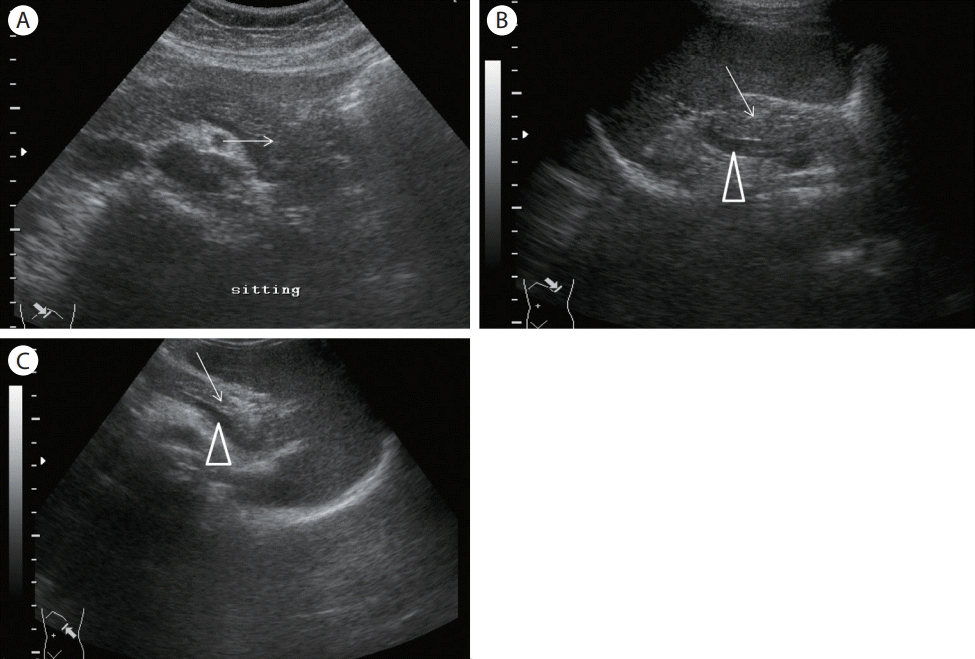
53세 여자로 타 병원에서 위장 질환으로 두 달간 치료하였으나 증세의 호전이 없어 내원하였다. 진료 후 초음파 검사하여 심와부 사 스캔과 경비장 스캔에서 이상이 없음을 확인하였으나, 우하측와위 좌늑골하 횡 스캔에서 췌장 미부에 저에코 종괴가 발견되었다(Fig. 2). 복부 전산화단층촬영상 췌장 미부암으로 진단되어(Fig. 3) 상급병원에서 수술하였다.

Ultrasound examination of case 1 (53/female, epigastric pain). (A) Epigastric transverse scan shows no remarkable findings. (B) Epigastric oblique scan also shows no remarkable findings. (C) Transsplenic scan fails to reveal the pancreas tail due to bowel gas. (D) Left subcostal transverse scan with a right lateral decubitus position demonstrates an oval hypoechoic mass in the tail of the pancreas (2 cm) (arrow).
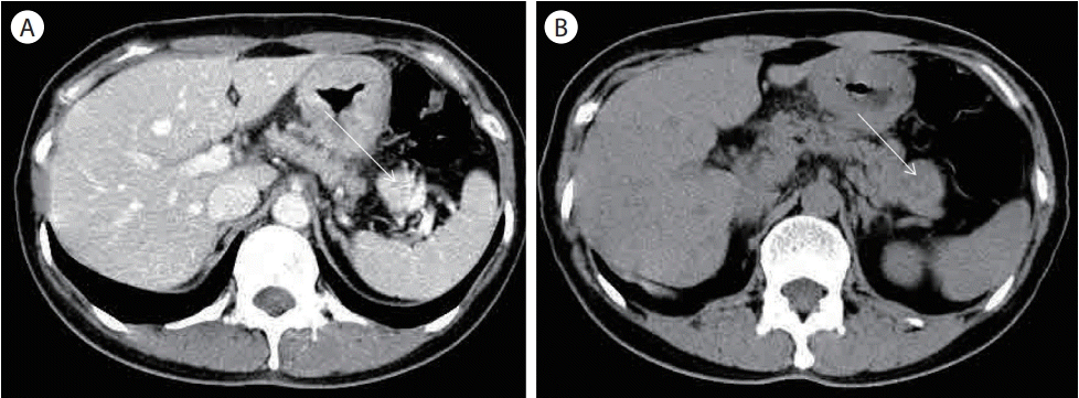
Abdominal computed tomography of case 1 (53/female, epigastric pain). (A) Contrast enhancing hypodense mass in the distal tail of the pancreas is suggestive of pancreatic cancer (arrow). (B) Hypodense mass in the tail of the pancreas (arrow).
증례2
73세 여자로 복통으로 본 의원에서 진료 후 초음파 검사를 하였다. 심와부 횡 스캔 및 사 스캔에서 췌장 체부 이하는 전혀 보이지 않아 우하측와위 좌늑골하 횡 스캔을 하였고 췌장 미부에 저에코 또는 무에코 종괴가 췌관과 연결되어 관찰되었다. 복부 전산화단층촬영상 분지형 췌관 내 유두상 점액종양으로 진단되었다(Fig. 4).
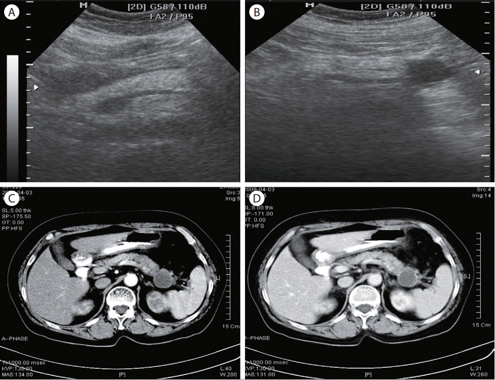
Ultrasound examination and abdominal computed tomography of case 2 (73/female, epigastric pain). (A) Epigastric transverse scan does not show the pancreas tail. (B) Left subcostal transverse scan with a right lateral decubitus position reveals an oval hypoechoic mass with a relatively smooth margin in the tail of the pancreas. The mass communicated with the pancreatic duct. (C, D) A hypodense mass with a faintly enhancing rim in the tail of the pancreas that communicates with the pancreatic duct. Branch duct-type intraductal papillary mucinous neoplasm is suspected.
증례3
58세 여자로 건강검진 목적으로 초음파 검사를 하였다. 심와부 사 스캔에서 이상이 없었으나 우하측와위 좌늑골하 횡 스캔에서 췌장 미부에 낭성 병변이 있고 낭종 벽에 석회화가 있어 점액성 낭성종양이 의심되었고 복부 전산화단층촬영에서 확인되었다(Fig. 5).
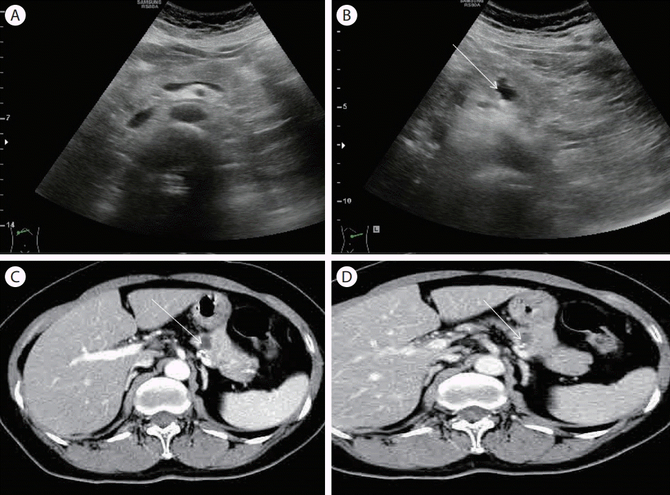
Ultrasound examination and abdominal computed tomography of case 3 (58/female, general medical examination). (A) Epigastric transverse scan does not show the pancreas tail. (B) Left subcostal transverse scan with a right lateral decubitus position shows an oval hypo- or anechoic cystic mass with a slightly irregular margin and wall calcifications (arrow). (C, D) A hypodense mass with wall calcifications and enhancement raises suspicions of mucinous cystic neoplasm (arrow).
결 론
췌장 초음파 검사는 췌장 질환 진단의 기본적인 검사이지만 그 해부학적 위치와 여러 방해인자 때문에 검사가 쉽지 않다[1-3]. 특히 췌장 미부는 관찰이 어려워 췌장 미부의 병변을 놓치는 경우가 적지 않다[1,3,4]. 췌장 미부 초음파 스캔법으로는 심와부 사 스캔, 경비장 스캔이 널리 이용되고 있으며 필요 시에는 음수법을 사용하기도 한다[1,4,5].
그러나 일반적인 췌장 미부 스캔법(심와부 사 스캔, 경비장 스캔)으로는 췌장 미부를 충분히 관찰할 수 없는 경우가 많아 췌장 미부 질환을 놓치는 경우가 많다. 특히 작은(2 cm 미만) 췌장 종양(암, 낭성 병변 등)인 경우는 더욱 관찰이 힘들다[5]. 물론 내시경 초음파나 복부 전산화단층촬영 등의 검사가 복부 초음파보다 췌장 미부 질환의 진단에 더 유용하다고 생각하지만 복부 초음파도 검사자의 노력에 따라 췌장 미부 질환을 상당수에서 찾아낼 수 있다고 생각한다. 저자는 췌장 미부 관찰을 위하여 초음파 검사 시 심와부 사 스캔, 경비장 스캔과 함께 우하측 와위 좌늑골하 횡 스캔을 시행하였다. 우하측와위 좌늑골하 횡 스캔으로 심와부 사 스캔이나 경비장 스캔에서 보이지 않았던 췌장 미부 질환을 다수 발견하였기에 췌장 미부 스캔시 심와부 사 스캔, 경비장 스캔과 함께 우하측와위 좌늑골하 스캔을 꼭 시행할 것을 권유한다.