갑상선염의 초음파 소견
Ultrasound Findings of Thyroiditis
Article information
Trans Abstract
Thyroiditis is a general term that refers to the inflammation of the thyroid gland. It can be classified as acute, subacute (painful and painless), or chronic. Clinical symptoms may include pain and tenderness when caused by infection, but it may be painless when caused by autoimmune conditions. It can be associated with normal, increased, or decreased thyroid function, often with changing from one condition to another. Several parameters evaluated by the ultrasound (US) contribute to the diagnosis. This article reviews the types of thyroiditis and their US findings.
서 론
갑상선염은 갑상선에 발생하는 염증성 질환으로 크게 급성, 아급성, 만성 갑상선염으로 분류하며, 원인은 세균, 바이러스 등의 감염에 의한 것과 자가면역에 의한 것 등으로 다양하다[1](Table 1). 갑상선은 초음파로 관찰하기 쉬운 장기로 초음파를 이용하면 다양한 갑상선 질환을 침상 옆에서 진단이 가능하다. 특히 갑상선의 병태생리를 잘 이해하고 있으면 형태학적 정보에 임상적인 소견을 더함으로써 보다 정확한 진단이 가능하다. 또한 컬러도플러를 이용하면 갑상선혈류 관찰로 갑상선기능을 예측할 수 있기 때문에 그레이브스병과 무통성 갑상선염을 감별할 수도 있다[2]. 본론에서는 임상에서 흔히 접하는 갑상선염의 초음파 소견에 대해 살펴보고자 한다.

Charcteristics of thyroiditis
본 론
급성 갑상선염(급성 화농성 갑상선염)
급성 화농성 갑상선염은 드문 질환으로 상기도염이나 중이염 등 선행하는 감염질환의 후유증으로 발생하거나, 양배꼴동루(pyriform sinus fistula)나 갑상설관 낭종(thyroglossalduct cyst) 등이 동반될 때 잘 발생하며, 면역이 저하된 환자에서도 발생한다. 감염이 원인이며, 결핵균, 살모넬라균, 방선균, 황색 포도상구균, 연쇄상구균, 폐렴구균 등의 세균에 의한 경우와 그 외 기생충 및 진균 감염에 의한 경우도 보고되어 있다. 급성 화농성 갑상선염은 전경부의 발적, 동통, 발열 등을 호소하고, 갑상선 기능검사는 대부분 정상이나, 아주 드물게 갑상선중독증이 동반된 예가 보고되어 있다[3]. 초음파 소견으로는 경계가 불명확한 저에코 음영을 보이며, 화농의 양이 많은 경우에는 내부에 부스러기를 동반한 낭종의 소견을 보인다(Fig. 1). 세침흡인세포 검사를 할 경우 배농이 되므로 진단이 가능하다.
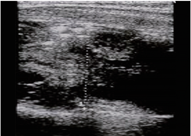
Ultrasonography of the neck reveals a left thyroid mass of about 2.4 cm with heterogeneously increased internal echogenicity. This mass was diagnosed as a thyroid abscess by a fine needle aspiration biopsy.
아급성 갑상선염
아급성 갑상선염은 압통을 동반하는 일과성 염증성 질환으로, 주로 상기도 감염 이후에 발현되는 경우가 많아 바이러스 감염이 원인으로 추정되고 있다[4]. 아급성 갑상선염의 임상경과는 갑상선 중독기와 회복기를 거쳐 대개 4-6개월 후에는 호전이 되나, 드물게 영구적인 기능저하증을 보이거나 재발하기도 한다. 아급성 갑상선염과 초기 그레이브스병과 감별진단은 적혈구 침강속도(erythrocyte sedimentation rate, ESR)의 증가, 갑상선 수용체항체 음성, 갑상선스캔과 24시간 방사성요오드 섭취율(radioactive iodine uptake, RAIU) 저하로 가능하다. 개원가에서는 핵의학적 검사가 가능하지 않는 경우가 많으므로 아급성 갑상선염의 진단은 먼저 문진과 진찰시 갑상선 부위에 압통이 있는지를 파악하는 것이 중요하며, ESR 검사와 갑상선 초음파검사로도 진단을 할 수 있다. 아급성 갑상선염의 전형적인 초음파 소견은 갑상선 한쪽 엽 또는 양엽에 경계가 불분명한 현저한 저에코 음영(또는 국소적 지도 모양 병변)을 보이는 것이다(Fig. 2).

Subacate thyroiditis ultrasound in transverse (A, C) and longitudinal (B, D) view shows a poorly defined hypoechoic nodular lesion in the thyroid lobe.
무통성 갑상선염
무통성 갑상선염은 임상경과는 아급성 갑상선염과 같으나 갑상선에 동통 및 압통이 없는 것이 다르며, 병리 소견상 림프구 침윤이 현저하고 혈액검사에서 갑상선 자가항체가 검출되는 경우가 많아 하시모토 갑상선염의 변형으로 생각되고 있다. 무통성 갑상선염은 산발적으로 발생하는 경우와 출산 후에 발생하는 경우가 있는데 후자를 산후 갑상선염이라고 한다. 무통성 갑상선염 초기에는 그레이브스병과 감별이 요하는데, 갑상선 수용체항체 음성, 갑상선스캔과 RAIU 저하를 확인함으로써 가능하다. 초음파검사 소견은 하시모토 갑상선염의 소견과 유사하며, 컬러도플러검사에서 혈류 증가가 없는 것으로 그레이브스병과 감별이 가능하다[5] (Fig. 3). 특히 산후에 모유 수유 중에는 RAIU를 시행할 수 없어 감별진단이 어려우나 컬러도플러는 태아에 해가 없으므로 아주 유용하다.

Painless thyroiditis. A transverse view shows diffuse enlargement, presenting reduced echogenicity and heterogeneous texture (A) and decreased parenchymal vascularization in the thyroid gland (B).
만성 갑상선염
만성 갑상선염에는 자가면역 국소 갑상선염, 만성 림프구성 갑상선염(하시모토 갑상선염), 위축성 갑상선염, 리델 갑상선염 등이 있으며, 이 중 하시모토 갑상선염이 가장 흔하다. 하시모토 갑상선염의 자가면역반응은 림프여포를 형성하는 림프구와 형질세포의 침윤을 일으켜, 대부분의 갑상선여포를 파괴하며, 점차적으로 섬유화가 진행되고 정상 갑상선이 림프여포와 섬유화로 대체되면서 갑상선 기능저하증을 초래한다[6]. 하시모토 갑상선염의 초음파 소견은 갑상선의 미만성 종대, 실질 에코의 저하, 미세결절화 및 거친 섬유성 중격 형성이 특징적이다[7] (Fig. 4). 갑상선의 림프구 침윤으로 정상 여포 구조가 파괴되고 세포 충실도가 높아지므로 실질 에코는 감소하며 소엽 구조가 과장되어 미세결절화가 발생한다. 갑상선혈류는 정상이거나 감소하지만 갑상선저하증이 있는 경우 그레이브스병에서 보이는 혈류 증가 소견이 보일 수 있다. 국소성 염증성 결절이 관찰되는 경우가 흔하지만 뚜렷하게 구별되는 결절성 병변이 보이는 경우 갑상선림프종 혹은 유두암종과의 감별이 요하기도 한다. 위축성 갑상선염은 갑상선자극호르몬 수용체 항체 중 억제작용을 하는 항체(blocking antibody)가 작용할 때 갑상선이 위축하게 되고 영구적인 기능저하증이 발생한다(Fig. 5).
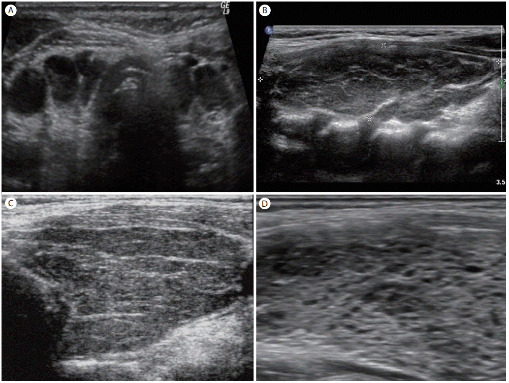
Hashimoto’s thyroiditis. Thyroid ultrasonography shows diffuse enlargement, hypoechogenicity, and heterogeneous texture with micronodulation in the thyroid gland (A-D).

Atrophic thyroiditis. Ultrasound shows a diffusely hypoechogenic thyroid gland with reduced-volume compared to the normal pattern.
결 론
초음파검사는 다른 검사에 비해 방사선의 피폭이 없고, 검사가 단순하고 비침습적이며, 반복하기 쉽고, 특히 침상 옆에서 즉시 정보를 얻을 수 장점이 있어 최근 갑상선질환의 감별진단에 흔히 이용되고 있다. 갑상선 결절의 감별진단에 비해 갑상선염의 진단에 있어 초음파검사의 역할은 제한적이지만, 임상증상과 혈액검사와 병행을 하면 큰 도움을 받을 수 있을 것으로 생각한다.