갑상선 유두암 초음파 소견과 병리학적 근거
Ultrasonographic Diagnosis of Thyroid Papillary Carcinoma with Pathologic Base
Article information
Trans Abstract
Ultrasonographic examination of the thyroid is simple and easy due to superficial anatomy and easy accessibillity. Although its anatomy is simple, the pathologic fingdings of thyroid diseases are quite diverse. Diagnosis and differentiation of thyroid ultrasonogrphic images are essential for pathologic knowledge. This review describe the sonographic findings associated with pathologic base.
서 론
갑상선은 표재성 장기로 고해상도 고주파 탐촉자를 이용하면 선명하고 자세한 영상을 얻을 수 있다. 갑상선질환에서 보이는 다양한 영상 소견을 이해하는데는 병리적 지식이 필요하다. 임상에서 흔히 접할 수 있는 갑상선 유두암의 초음파 소견과 병리적 배경에 대해서 간략하게 기술한다.
본 론
전형적인 유두암(classical papillary carcinoma)
병리 조직 소견(Figs. 1-3)
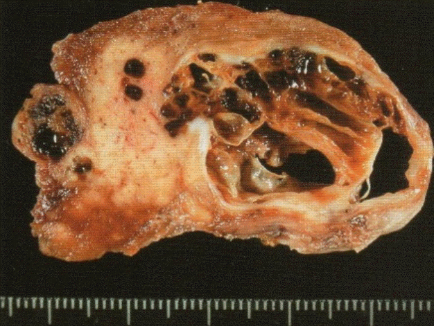
Cystic papillary carcinoma. Surgical specimen showing solid tumor mass with multiple cystic components.
유두상 구조를 기본으로 하지만 일반적으로 여포 구조를 나타내는 부분과 혼재되어 있다.
종양세포는 압방형 또는 원주상이고 호산성 색조로 보인다.
핵은 인접하는 핵과 중첩되고 모양은 유원형이지만 초생달 형태도 관찰된다.
핵의 chromatin은 미세 과립상으로 소위 ground glass 형태를 보이기도 한다.
핵의 장축에 평행한 방향으로 생기는 핵구(nuclear groove)와 세포질 함입으로 가성 핵내 세포질 봉입체(nuclea r pseudoinclusion)가 특징적인 소견이다.
때로는 사립 소체가 보이고 이는 유두상 구조에서 간질 부위의 석회화이다.
편평 상피 화생도 보이나 이는 예후와 관계없다.
초음파 소견과 병리학적 근거
• 고분화형 유두암은 초음파상 저에코 또는 현저한 저에코 결절로 보인다.
⇒ 여포 상피의 증식으로 주위 여포조직을 파괴하여 콜로이드가 감소되고 암성 증식된 세포들의 초음파 투과성 항진으로 저에코로 보인다.
• 종괴의 경계는 불규칙하고 주위 조직으로 침윤하는 모습을 보인다.
⇒ 유두암이 주위 조직을 침범하므로 결절의 경계가 불규칙하게 보인다.
• 종괴의 내부에 점상 석회화 소견을 보인다.
⇒ 유두암에서 유두상 구조의 간질의 석회화가 사립 소체(psammoma body)이고 크기가 1 mm 이하이면 후방 음영을 동반하지 않는다.
• 초음파 횡단상에서 결절이 앞뒤로 길쭉한 형태를 보인다.
⇒ 양성 결절과 달리 성장 방향이 주변 조직에 관계없이 진행되어 소위 taller than wide 모양을 보이기도 한다.
• 유두암도 낭성 변성을 일으키나 완전한 낭성 변성은 드물다.
⇒ 초음파 검사시 고형 성분의 형태, 에코, 내부의 미세 석회화 및 주위 실질 침범 여부를 관찰해야 한다.
• 경부 림프절 종대가 흔히 동반되고 림프절 내부에 미세 석회화, 낭성 변성이 보이면 전이를 강력히 시사한다.
⇒ 유두암은 림프절 전이를 잘 일으키고 초음파상 보이지 않는 작은 유두암에서도 인접한 경부 림프절에 전이를 동반하는 경우도 있다.
Case 1 (Figs. 4-7)
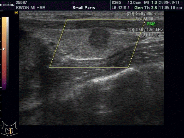
Ultrasonogram of showing a marked hypoechoic nodule with lobulated margin, multiple microcalcifications.
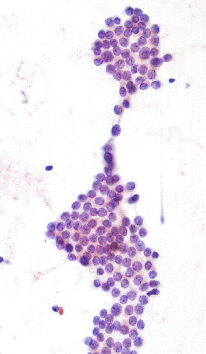
Papillary carcinoma. Microscopic finding showing papillary carcinoma features. Pap, smear 400x magnification.

Papillary carcinoma. Microscopic finding showing papillary carcinoma features (fine chromatin, irregular nuclear margin, dense cyoplasm). Pap, smear 400x magnification.
현저한 저에코 분엽상을 보이며 내부 미세 석회화상을 동반한 좌측 갑상선 유두암(F/32). 32세 여성으로 건강검진에서 발견된 좌측 갑상선 결절의 세포진을 위해 내원함. 내원시 vital signs은 정상이고 갑상선기능 이상 증세는 없음.
세포진 소견
[Diagnosis]
Thyroid gland, left, aspiration cytology;
1) 1st fine needle aspiration (FNA) (1-3): Papillary carcinoma, consistent with
2) 2nd FNA (4-6): Papillary carcinoma, consistent with 수술을 시행하였다.
[Diagnosis]
Thyroid gland, both lobe, total thyroidectomy
(1) Right lobe: No pathologic abnormality
(2) Left lobe: papillary carcinoma, microcarcinoma variant with 1) tumor invasion into extrathyroid soft tissue
2) tumor size: 0.5 × 0.5 cm
3) vascular invasion (-)
4) surgical margin: tumor extension in the resecction margin
5) no-neoplastic thyroid: no pathologic abnormality surgical Pathological diagnosis: Left thyroid Papillary carcinoma
Case 2 (Figs. 8-11)
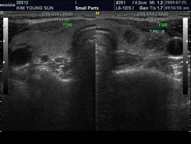
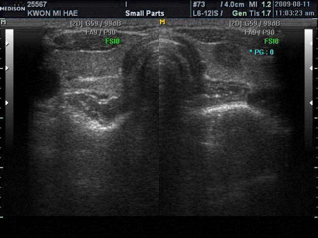
Both thyroid longitudinal scan. Ultrasonogram showing two hypoechoic nodule with irregular margin in the left thyroid.
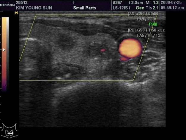
Left thyroid, longitudinal scan, power Doppler. Ultrasonogram showing no increased blood flow in the hypoechoic nodules.
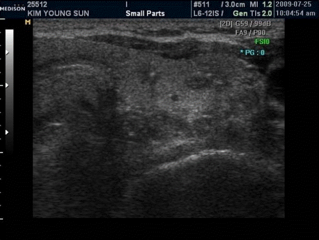
FNA left thyroid. Ultrasonogram showing needle tip in the medial side of the left thyroid nodule. FNA, fine needle aspiration.
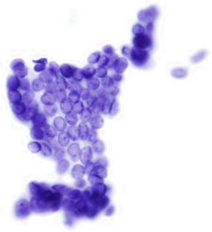
Papillary carcinoma. Cytological finding showing papillary carcinoma features (fine nuclear chromatins, pseudoinclusion, nuclear groove) Pap, smear. 400x magnification.
경계가 분명하고 분엽상을 보이는 현저한 저에코 결절로 관찰되는 좌측 갑상선 다발성 유두암(F/35). 35세 여성으로 건강검진에서 발견된 갑상선 결절의 세포진을 위해 본의원을 방문함. 내원시 vital signs은 정상이고 이학적 검사상 특이소견이 없고 갑상선기능 이상 증세는 없음.
세포진 소견
[Diagnosis]
Thyroid gland, l eft ( 1) m edial, a spiration c ytology ( 3): Papillary carcinoma, consistent with
Thyroid gland, l eft ( 2) l ateral, a spiration c ytology ( 3): Papillary carcinoma, consistent with
Total Thyroidoctomy
Surgical pathology report: Papillary cancer
Case 3 (Figs. 12-20)

Both thyroid, transverse scan. Ultrasonogram showing hypoechoic nodule with cystic component in the right thyroid.
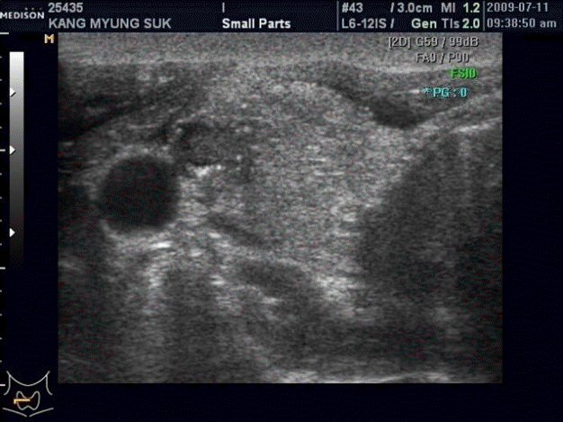
Right Thyroid, transverse scan. Ultrasonogram showing hypoechoic nodule with cystic component in the right thyroid.

Right thyroid, longitudinal scan. Ultrasonogram showing hypoechoic nodule with cystic area in the right thyroid.
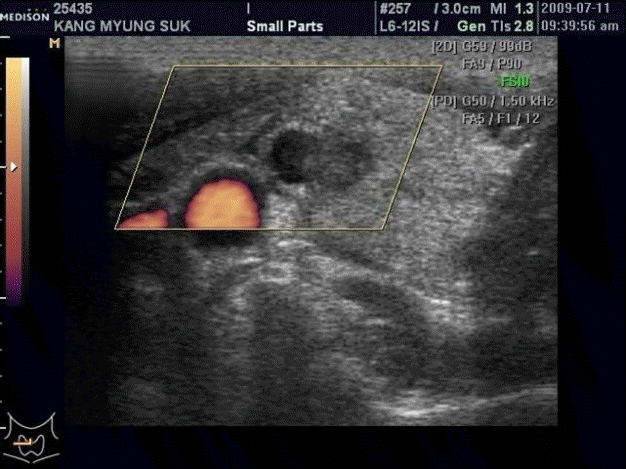
Right thyroid, transverse scan, power Doppler. Ultrasonogram showing no increased blood flow signal in the nodule.
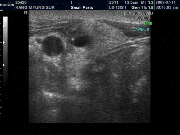
First FNA, right thyroid. Ultrasonogram showing needle tip in the cystic area of nodule. FNA, fine needle aspiration.

Second FNA, right thyroid. Ultrasonogram showing needle tip in the solid area of nodule. FNA, fine needle aspiration.
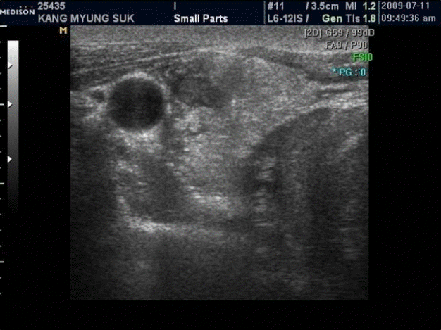
Third FNA, right thyroid. Ultrasonogram showing needle tip in the peripheral area of nodule. FNA, fine needle aspiration.
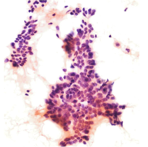
Papillary carcinoma. Cytological finding showing dome shaped structure, probably representing the tip of papilla. Pap, smear 400x magnification.
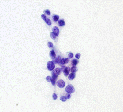
Papillary carcinoma. Microscopic finding of second FNA showing atypical follicular cells, favor neoplastic change. Pap, smear 100x magnification. FNA, fine needle aspiration.
결절의 변연부위 세포진으로 진단된 낭성 유두암(M/32). 32세 남성으로 건강검진에 발견된 우측 갑상선 결절의 세포진을 위해 본원을 방문함. 내원시 vital signs은 정상이고 갑상선기능 증세는 없음.
세포진 소견
[Diagnosis]
Thyroid gland, right, aspiration cytology:
1) 1st FNA (1-2): Rare follicular cells
2) 2nd FNA (3-4): Atypical follicular cells
3) 3rd FNA (5-6): Papillary carcinoma, consistent with
Total thyroidectomy with radical neck dissection을 시행하였다. Surgical pathologic diagnosis: Thyroid cancer with neck metastasis
Case 4 (Figs. 21-23)
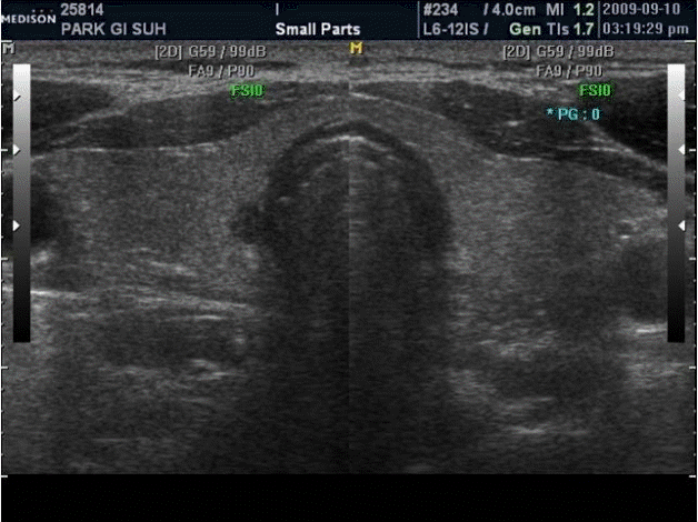
Both thyroid, transverse scan. Ultrasonogram showing hypoechoic nodule near the trachea in the right thyroid.
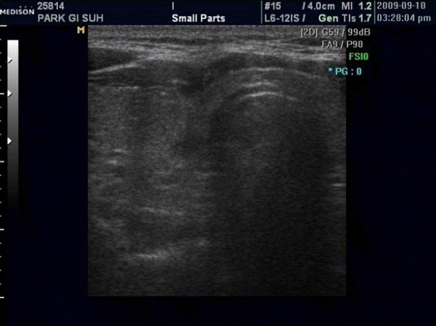
FNA right thyroid. Ultrasonogram showing needle tip in the right thyroid nodule. FNA, fine needle aspiration.
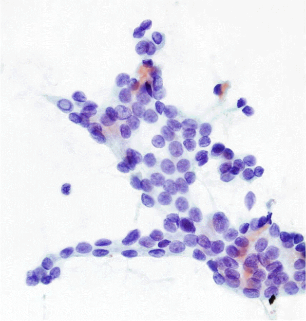
Papillary carcinoma. Microscopic finding showing papilary carcinoma feature (pseudoinclusion, fine nuclear chromatins, nuclear groove), Pap, smear 400x magnification.
기관옆에 인접하고 현저한 저에코상을 보이는 우측 갑상선 미소 유두암(M/42). 42세 남성으로 건강검진에서 발견된 우측 갑상선 결절의 세포진을 위해 본 의원을 방문함. 내원시 vital signs은 정상이고 이학적 검사상 특이소견은 없고 갑상선기능 이상 증세는 없음.
세포진 소견
[Diagnosis]
Thyroid gland, right, aspiration cytology:
1) 1st FNA (1-3): Papillary carcinoma, consistent with
2) 2nd FNA (4-6): Rare follicular cells
Right lovectomy를 시행하였다.
Surgical pathologic diagnosis: Thyroid papillary cancer, right lobe, 4 cm diameter
여포형 유두암(follicular variant of papillary carcinoma)
병리 조직 소견
종양이 전체적으로 여포상 구조로 구성되고 유두상 구조가 결핍된 유두암이다.
핵 소견으로 유두암을 진단하고 임상적으로 고분화형 유두암과 차이가 없다.
종양은 소-중형 크기의 여포가 서로 압박하는 형태로 밀집하여 증식한다.
종양의 경계는 피막으로 덮여 있는 경우가 많고 약확대상에서 여포성 종양으로 의심되지만 여포성 종양보다 핵의 직경이 크고 맑게 보이면 여포형 유두암을 의심할 수 있다.
핵 소견은 고분화형 유두암과 동일하고 ground glass 핵, 핵구, 핵내 세포질 가성 봉입체가 보인다.
사립 소체는 거의 보이지 않는다.
초음파 소견과 병리학적 근거
• 초음파상으로 양성 선종양결절, 여포성 종양과 감별이 곤란하다.
⇒ 결절의 크기가 1 cm 이상이면 여포성 종양, 여포형 유두암, 양성 선종양결절과의 감별을 위해 FNA을 권유하는 것이 좋다고 생각된다.
Case 5 (Figs. 24-27)

Both thyroid, transverse scan. Ultrasonogram showing round isoechoic nodule with peripheral halo in the right thyroid.
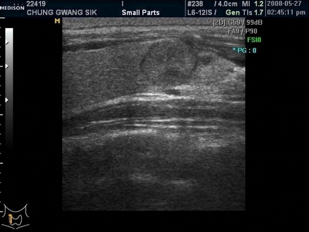
Right thyroid, longitudinal scan. Ultrasonogram showing round isoechoic nodule with cystic area in the right thyroid.

Right thyroid, transverse scan, color Doppler. Ultrasonogram showing no increased blood flow in the right thyroid nodule.
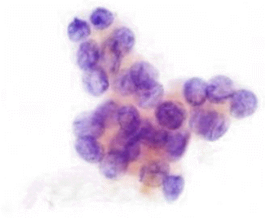
Papillary carcinoma. Cytological finding showing papillary carcinoma features. Pap, smear. 400x magnification.
초음파상 양성 결절상으로 보이는 우측 갑상선 유두암(M/34). 34세 남성으로 screening 초음파 검사에서 우측 갑상선결절 발견함. 내원시 vital sign은 정상이고 이학적 검사상 특이소견 없음.
세포진 소견
[Diagnosis]
Thyroid gland, right, aspiration cytology:
1) 1st FNA (1-3): Rare follicular cells, some blood smeared
2) 2nd FNA (4-6): Papillary carcinoma
결 론
갑상선 유두암은 임상에서 흔히 접하는 종양이다. 갑상선 유두암의 병리 지식을 기초로 초음파검사를 시행하면 진단에 큰 도움이 된다고 생각한다.
