어깨 질환의 초음파 소견
Shoulder Ultrasonography: Common Findings
Article information
Trans Abstract
Shoulder pain is one of the most common musculoskeletal complaints in clinical practice. Ultrasonography is a useful method for the diagnosis of various shoulder diseases and injuries. This article presents the ultrasonographic features of common shoulder conditions, including those affecting the rotator cuff, long head of the biceps, subacromial-subdeltoid bursa, and glenohumeral and acromioclavicular joints.
서 론
어깨 통증의 유병률은 15-20%에 이를 정도로 흔한 근골격계 증상이며, 특히 최근 노인층의 증가로 인하여 발생빈도가 증가하고 있다[1]. 어깨의 상완와관절(glenohumeral joint)은 운동범위는 넓지만 불안정한 관절 구조로 회전근개(rotator cuff)와 상완이두장두건(long head of the biceps brachii tendon)이 관절의 안정성 유지에 중요한 역할을 한다. 어깨 질환은 관절 내 문제보다는 회전근개를 비롯한 관절 주위 힘줄이나 윤활낭 등의 연부조직 질환에 의한 경우가 더 흔하다[2]. 특히 초음파 검사는 어깨 질환 진단에 있어서 자기공명영상과 비교하여 회전근개, 견봉하-삼각근하 윤활낭(subacromial-subdeltoid bursa), 상완이두장두건의 퇴행성 및 염증성 질환 등의 진단 정확도가 비슷하다. 그러나 자기공명영상과 비교하여 짧은 검사시간, 저렴한 비용 및 역동적 검사를 실시할 수 있다는 장점이 있어, 어깨 초음파는 근골격계 초음파 분야에서 가장 많이 이용되고 있다[3]. 본론에서는 임상에서 흔히 접하는 어깨 질환의 초음파 소견에 대해 살펴보고자 한다.
본 론
상완이두장두건 질환
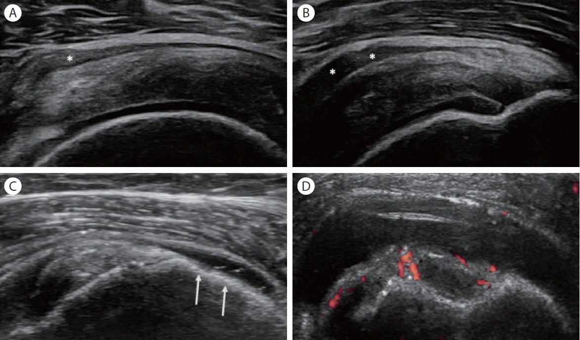
상완이두장두건은 상완와관절의 윤활막이 연장된 건초(tendon sheath)로 둘러 쌓여 있으며, 상완와관절과 연결되어 있기 때문에 정상에서도 소량의 활액이 힘줄 주위에 보일 수 있다[4]. 따라서 상완이두장두건 주위에 삼출액이 증가되면 힘줄 자체의 병변보다는 상완와관절의 질환을 먼저 고려해야 한다. 상완이두장두건의 건초염(tenosynovitis)이 있으면 건초가 국소적으로 확장되면서 혈류의 증가 소견을 보이며, 탐촉자로 누르면 통증이 동반될 수 있다(Fig. 1). 상완이두장두건의 건초염은 만성적인 반복적 충돌과 마모에 의해서 발생하며, 류마티스 관절염과 같은 염증성 관절염 및 감염성 관절염으로 인하여도 발생할 수 있다.

Tenosynovitis of the long head of the biceps tendon. (A) Transverse and (B) longitudinal ultrasonographic (US) images demonstrate hypoechoic hypertrophy and effusion (arrows) within the tendon sheath. The tendon sheath exhibits increased power Doppler signal on (C) transverse and (D) longitudinal US images.
상완이두장두건의 힘줄증(tendinosis)은 원발성으로 발생하는 경우는 드물다. 상완이두장두건의 힘줄증의 원인은 일반적으로 힘줄의 만성 반복적 사용과 관련된 견인(traction) 및 마찰력(friction forces)이다. 초음파에서 상완이두장두건 힘줄증은 힘줄의 두께가 두꺼워지고 저에코(hypoechoic)로 보인다. 상완이 두장두건의 근위부 파열은 기저의 힘줄증이나 퇴행성 변화로 인하여 발생하는 경우가 흔하다. 상완이두장두건의 전층 파열이 발생하면 원위부 힘줄의 뒤당김(retraction)으로 특징적인 “pop-eye” 징후를 보인다. 초음파에서 상완이두장두건의 파열은 힘줄이 보이지 않거나 결간구(bicipital groove)가 비어 있는 상태로 보인다.
견봉하-삼각근하 윤활낭 질환
정상 견봉하-삼각근하 윤활낭은 고에코로 보이는 두 층의 윤활낭 주위 지방 사이에 저에코 띠로 보이며, 정상 두께는 약 0.5 mm이며, 2 mm까지 정상 두께로 간주한다. 견봉하-삼각근하 윤활낭에 영향을 미칠 수 있는 질환에는 회전근개 질환에 이차적인 삼출(effusion), 감염성 또는 염증성 견봉하-삼각근하 윤활낭염(bursitis)이 포함된다. 회전근개의 전층 파열이 생기면 관절강(joint cavity)과 견봉하-삼각근하 윤활낭이 연결되면서 bursal distension이 발생한다[3]. 초음파에서 견봉하-삼각근하 윤활낭에 활액이 증가되면 무에코(anechoic) 혹은 저에코로 관찰되며 때때로 출혈, 감염 등으로 인하여 복합 에코로 보이기도 한다(Fig. 2A, B). 견봉하-삼각근하 윤활낭의 삼출액의 양이 많은 경우 낮은 곳으로 이동하므로, 대결절(greater tuberosity)의 바깥쪽까지 연장되어 눈물방울징후(tear-drop sign)로 관찰된다(Fig. 2C). 비후된 견봉하-삼각근하 윤활낭은 삼각근과 비슷한 에코로 관찰되기 때문에, 힘줄 또는 근육의 일부로 오인할 수 있다. 비후된 윤활낭에 가는 섬유다발 모양이 보이지 않는 것이 힘줄과의 감별점이다. 윤활낭 내의 활액 양이 증가하거나 활액막 비후 소견이 보이면, 류마티스 관절염, 통풍관절염과 같은 결정 유발성 관절염, 출혈, 감염성 윤활낭염 등의 원인도 함께 감별해야 한다(Fig. 2D) [5].
Subacromial-subdeltoid bursitis. Ultrasonographic (US) images of the supraspinatus on (A) transverse and (B) longitudinal views show hypoechoic bursal thickening (asterisks). US images of the supraspinatus on (C) longitudinal view show a tear-drop sign (arrows). Transverse US (D) demonstrates well-defined hypoechoic fluid accumulation and increased vascularity in the bursal space between the biceps tendon's long head and the deltoid muscle.
회전근개 질환
회전근개는 견갑하근(subscapularis), 극상근(supraspinatus), 극하근(infraspinatus), 소원근(teres minor)과 그 힘줄로 구성되며 어깨관절의 안정성과 움직임을 제공한다. 회전근개 질환은 가장 흔한 어깨 질환이며, 회전근개 힘줄병증(tendinopathy)과 파열이 흔하다[6].
회전근개 힘줄병증과 파열(rotator cuff tendinopathy and tear)
회전근개 힘줄병증의 초음파 소견은 다른 힘줄병증과 마찬가지로 힘줄의 비후와 함께, 힘줄이 국소적 혹은 전반적으로 저에코성으로 관찰되며, 정상적인 원섬유구조(fibrillary structure)가 소실되어 보인다(Fig. 3) [7]. 힘줄에 국소 병변이 확인되면 장축 및 단축 영상에서 모두 병변을 확인해야 하고, 비등방성(anisotropy)과 구분하기 위하여 탐촉자의 각도를 조절하는 것이 중요하다[8]. 힘줄의 주행 방향이 구부러지는 경우에는 비등방성에 의하여 에코가 감소하므로, 이 부분을 힘줄병증이나 파열로 오인하지 않는 것이 중요하다. 힘줄병증의 초음파 소견이 경미한 경우에는 무증상인 반대편 어깨와 비교해보는 것이 도움이 된다.
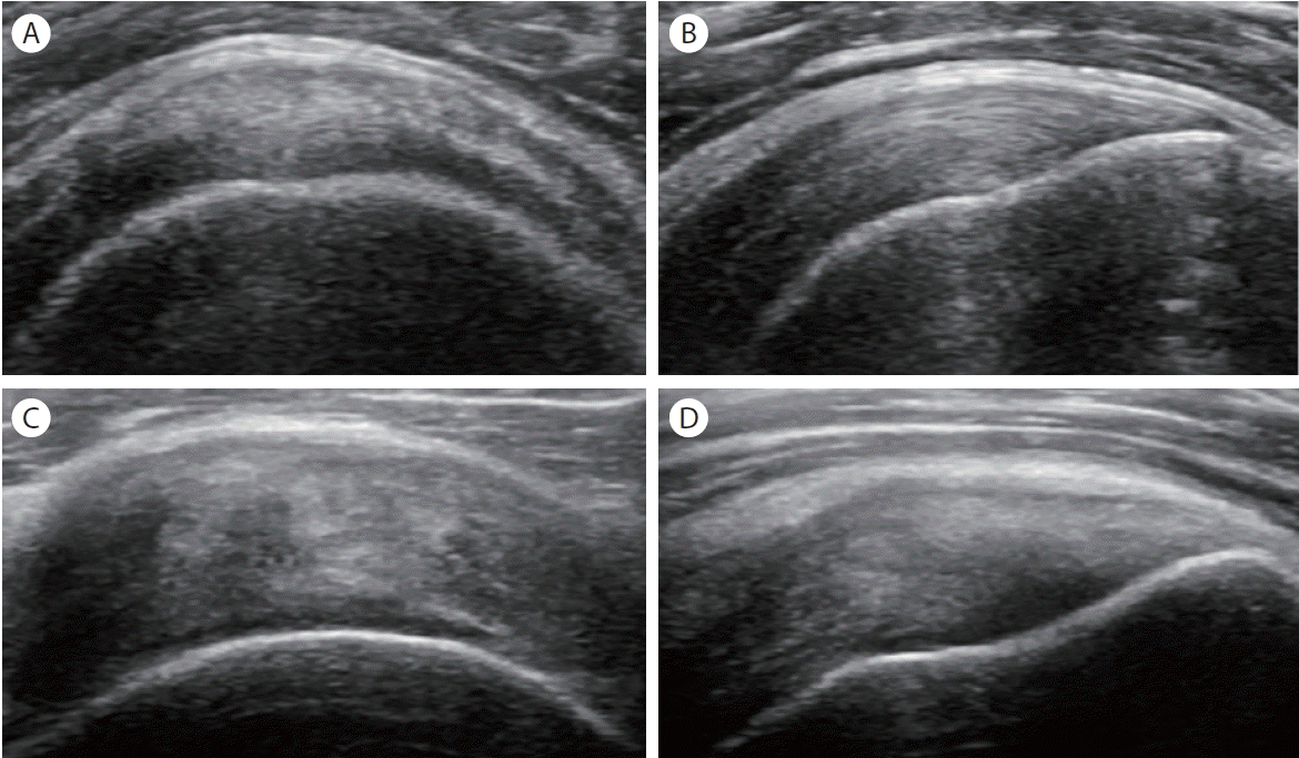
Tendinosis of the supraspinatus tendon. In contrast to normal supraspinatus tendon on (A) transverse and (B) longitudinal ultrasonographic (US) images, (C) transverse and (D) longitudinal US images of tendinosis show heterogeneous echogenicity, increased thickness and loss of fibrillar pattern of the tendon.
회전근개 파열은 극상근 힘줄에서 가장 많이 발생한다. 회전근개 파열은 전층 파열(full-thickness tear)과 부분층 파열(partial thickness tear)로 구분한다. 전층 파열은 힘줄의 관절면(articular surface)부터 윤활낭면(bursal surface)까지 힘줄의 전층을 침범하는 파열이다. 전층 파열은 힘줄 너비(width)의 일부를 침범하는 불완전(incomplete) 파열과 힘줄의 전체 너비를 침범하는 완전(complete) 파열로 나뉜다. 부분층 파열은 힘줄 두께의 일부분에 국한된 파열로, 파열 부위에 따라 윤활낭면, 관절면, 힘줄내(intrasubstance) 파열로 구분한다[3,6].
초음파 검사에서 전층 파열은 파열 부위의 저에코 혹은 무에코의 힘줄 결손(defect)으로 관찰된다(Fig. 4A, B). 특히, 회전근개 힘줄의 급성 전층 파열은 힘줄의 중간 부위에서 많이 발생하고, 파열 부위가 활액으로 차 있으며 관절이나 윤활낭의 삼출 소견이 관찰되는 경우가 많다. 만성 전층 파열은 힘줄의 뒤당김이 발생하여 힘줄이 보이지 않고, 상완골두가 힘줄로 덮이지 않아, 위쪽 삼각근이나 윤활낭 주위 지방에 맞닿아 상완골두가 노출되는 naked head sign을 볼 수 있다[3,6]. 회전근개 파열의 이차적인 간접적인 징후로는 대결절의 불규칙한 피질(cortical irregularity), 저에코의 두 구조물인 파열된 힘줄과 상완 골두 유리질 연골의 사이 경계면에서 고에코 선이 보이는 연골경계면징후(cartilage interface sign), 상완와관절의 삼출, 삼각근과 윤활낭 주위 지방이 파열 부위 내로 밀려 내려오는 sagging peribursal fat sign 등이 있다(Fig. 4C, D).
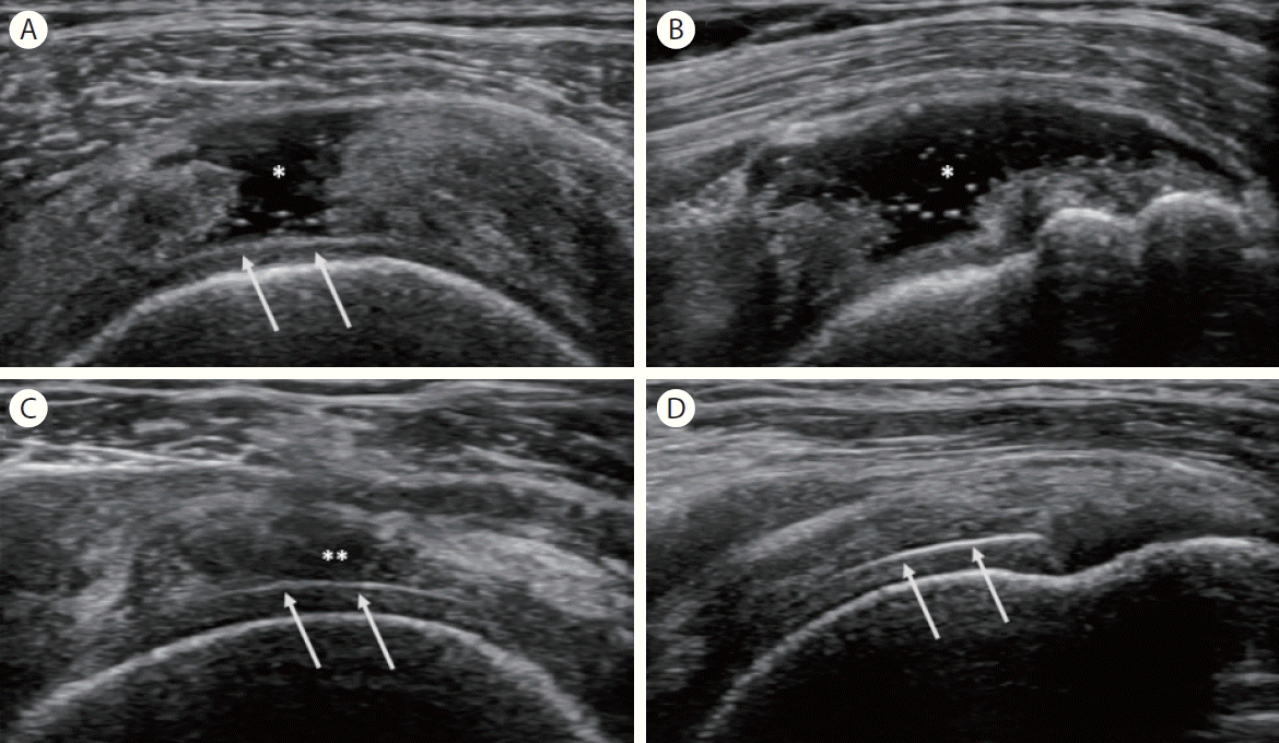
Rotator cuff tear. Ultrasonographic (US) images of the supraspinatus on (A) transverse and (B) longitudinal views show a fluid-filled anechoic defect (asterisk) and cartilage interface sign (arrows). US images of the supraspinatus tendon on (C) transverse and (D) longitudinal views show a hypoechoic defect (double asterisk) on the articular surface and cartilage interface sign (arrows).
석회성 건염(calcific tendinitis)
석회성 건염은 힘줄 내에 칼슘이 침착되는 질환으로 대부분 칼슘수산화인회석(calcium hydroxyapatite crystal)이 침착된다. 석회성 건염은 30대에서 50대에 호발하며 회전근개 중 극상근 힘줄에 가장 많이 발생한다. 석회성 건염의 원인은 명확하지 않지만, 퇴행성 변화, 대사성 요인, 유전 소인 등 다양한 요소가 작용하는 것으로 알려져 있다. 석회성 건염은 석회화 전 단계(pre-calcific), 석회화 단계(형성기와 흡수기), 석회화 후 단계(post-calcific)로 구분하며, 대개 흡수기(resorptive phase)에 심한 통증을 유발한다. 초음파 검사는 석회 침착의 특징적 소견과 위치를 파악하는 데 유용하다[9]. 회전근개의 석회는 힘줄 내의 경계가 불명확한 양털 모양 혹은 경계가 뚜렷한 고에코성 침착(well-defined hyperechoic deposits)으로 관찰되며, 소리그림자(acoustic shadowing)가 보이거나 보이지 않을 수 있다(Fig. 5). 도플러 검사는 흡수기와 형성기를 구분하는 데 도움이 되며, 흡수기에는 도플러가 검사에서 혈류 증가 소견을 보이는 경우가 많다[3].
Calcific tendinitis of the supraspinatus tendon. (A) Transverse and (B) longitudinal ultrasonographic images show a hyperechoic linear lesion (arrows) with posterior acoustic shadowing in the tendon.
상완와관절과 견봉쇄골관절 질환
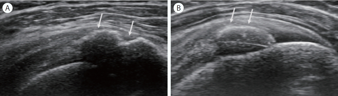
상완와관절의 퇴행성 관절염은 초음파 검사에서 좁은 관절 간격(joint space narrowing), 골극(osteophyte), 불규칙한 피질골(cortical irregularity)로 관찰된다. 상완와관절의 퇴행성 관절염은 회전근개의 만성 파열과 연관된 경우가 많다[6]. 견봉쇄골관절(acromioclavicular joint)에서 가장 흔한 병변은 퇴행성 관절염이다. 초음파에서 관절낭이 확장되며, 상완와관절과 마찬가지로 좁은 관절 간격, 골극, 불규칙한 피질골이 관찰된다(Fig. 6). 초음파 검사에서 관절 간격이 넓어진 소견을 보이면 외상, 감염성 또는 염증성 질환을 고려해야 한다. 류마티스 관절염은 어깨의 상완와관절과 견봉쇄골관절을 모두 침범할 수 있다. 류마티스 관절염의 활막염(synovitis)은 초음파 검사에서 활막 비후(synovial hypertrophy), 삼출(effusion), 도플러 검사에서 혈류 증가로 관찰된다. 특히 도플러 검사는 류마티스 관절염과 같은 염증성 질환과 퇴행성 질환의 감별에 도움이 될 수 있다(Fig. 7) [3].
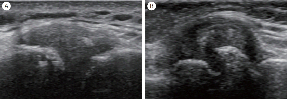
Osteoarthritis of the acromioclavicular joint. In contrast to the normal acromioclavicular joint seen on (A) long-axis ultrasonographic (US) images, the osteoarthritic joint shows osteophytes, joint space narrowing, and capsular distension on (B) long-axis US images in a patient with osteoarthritis.
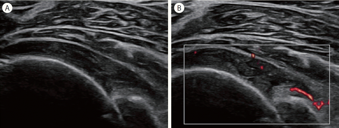
Synovitis of the glenohumeral joint. Transverse ultrasonographic (US) imaging of the posterior glenohumeral joint shows (A) joint effusion combined with hyperechoic synovial proliferation and (B) increased vascularity on power Doppler US.
결 론
어깨 초음파는 근골격계 초음파 검사에서 가장 많이 이용하는 분야 중 하나이다. 어깨의 해부학적 지식과 함께 각 질환에 대한 이해가 선행된다면, 임상에서 초음파 검사를 활용하여 다양한 회전근개 및 회전근개외 질환의 진단과 치료에 활용할 수 있을 것이다.