갑상선 고형 변이종 유두암의 초음파와 임상적 특징
Ultrasonographic and Clinical Characteristics of the Solid Varient of Papillary Thyroid Carcinoma
Article information
Abstract
목적
고형 변이종 유두암은 갑상선 유두암의 드문 변이종으로 임상적, 초음파적 특성이 잘 알려져 있지 않다. 본 연구는 조직학적으로 확진된 17예의 고형 변이종 유두암의 임상적, 초음파적 특성을 알아보고자 시행하였다.
방법
2009년 1월부터 2013년 9월까지 고신대학교 복음병원에서 갑상선절제술을 받은 환자 중 갑상선 유두암으로 진단된 4,136명 중 고형 변이종 유두암으로 진단된 환자 17예(0.41%)를 대상으로 후향적으로 연구를 시행하였다.
결과
고형 변이종 유두암 17예 중 남자는 2예(11.7%), 여자 15예 (88.3%)였으며, 평균 연령은 49.2세(범위 19-71세)였다. 종양의 크기는 평균 1.4 ± 0.6 cm (범위 0.5-3.2 cm)였다. 림프절 전이는 10예(58.9%)에서 관찰되었고, 그중 9예(52.9%)는 중심림프절 전이를, 5예(29.4%)는 외측림프절 전이를 보였다. 12예(70.6%)에서 병리학적으로 갑상선외 침범을 보였다. 평균 추적 기간은 33.5 개월(범위 6-67 개월)이었고, 재발한 예는 없었다. 초음파검사에서 결절의 모양은 난원형(ovoid)이 3예(17.6%)였고, 불규칙형 (irregular) 8예(47.1%), 앞뒤로 긴 모양(taller-than-wide shape)이 6예(35.3%)였다. 에코 특성은 등에코(isoechoic) 1예(5.9%)를 제외하면 나머지 16예(94.1%) 모두 저에코(hypoechoic) 또한 현저한 저에코(markedly hypoechoic)를 보였다. 결절의 변연부는 경계가 분명한(well defined) 결절이 1예(5.9%)였고, 나머지 결절은 경계가 불분명(ill defined)하거나 침상(spiculated) 경계를 보였다. 결절의 조성은 혼합성(mixed) 1예(5.9%)를 제외하고 16예(94.1%) 모두 고형성(solid)이었다. 미세석회화는 7예(41.1%)에서 관찰되었다.
결론
본 연구를 통하여 고형 변이종 유두암이 원격전이와 재발이 없어 공격적이지 않음을 알 수 있었다. 또한 고형 변이종 유두암은 초음파검사에서 앞뒤로 긴 모양, 현저한 저에코, 침상 변연부, 미세석회화 등의 악성을 시사하는 특징들을 보이는 경향이 있었다.
Trans Abstract
Background/Aims
The solid variant of papillary thyroid carcinoma (SVPTC) is one of rarest subtype of papillary thyroid carcinoma. There are few studies of the clinical and ulrasonographic (US) features of SVPTC. The aim of this study was to evaluate the clinical and ultrasonographic features of patients with 17 SVPTC treated at our institution.
Methods
Of the 4,136 patients with PTC who were underwent thyroidectomy at our institution between January 2009 and September 2013, 17 patients with SVPTC were retrospectively reviewed.
Results
Of those 17 patients with SVPTC, two (11.7%) were men and 15 (88.3%) were women, with a mean age of 49.2 years (range: 19-71 years). The mean tumor size was 1.4 ± 0.6 cm (range: 0.5-3.2 cm). The lymph node metastases were observed in 10 patients (58.9%). The extrathyroidal invasions were found in twelve (70.6%). The mean follow-up period was 33.5 months (range: 6-67 months). No patient experienced tumour recurrence or distant metastasis during follow-up. On US, the majority of nodules had irregular (8 [47.1%]) or taller than wide (6 [35.3%]) shapes with solid (16 [94.1%]). The margin of nodules was ill-defined or spiculate margins (16 [94.1%]). The echogenecity of nodules was hypoechoic (7 [41.2%]) or markedly hypoechoic (9 [52.9%]). The microcalcification was seen in seven (41.1%).
Conclusions
The SVPTC is not associated with distant metastasis and poor prognosis as previously thought. The SVPTC tends to have relatively malignant sonographic features such as taller than wide shape, markedly hypoechoic, spiculated margin and microcalcifications.
서 론
갑상선 유두암은 갑상선암 중 가장 흔하며 모든 갑상선암의 70% 이상을 차지한다[1]. 유두암 중에는 유두암의 특징적인 세포 형태를 보이는 고전적(classic) 유두암과 특징적 세포형태를 보이지 않는 변이종이 있으며, 변이종 중에는 여포성 변이종(follicular variant), 고형(solid) 변이종, 종주형(trabecular) 변이종, 인슐라형(insular) 변이종, 원주형 세포(columnar cell) 변이종, 키큰 세포(tall cell) 변이종, 체모양-오디모양(cribriform-morular) 변이종, 미만성 경화형(diffuse sclerosing) 변이종 등이 알려져 있다[2-4]. 여포성 변이종은 흔하고 예후가 양호하나, 그 외 변이종은 드물고 예후가 고전적 유두암보다 불량한 것으로 알려져 유두암의 침습형 변이종(aggressive variant of papillary carcinoma)이라고 한다[5,6].
고형 변이종 유두암은 1985년 Carcangiu 등[7]에 의해 처음으로 기술되었으며, 유두암 중 3%를 차지하는 드문 종양으로 생물학적 특성은 잘 알려져 있지 않으며[8], 체르노빌 원전사고 후 방사선에 노출된 소아에서 흔히 관찰되어 방사선 노출과 관련이 높은 것으로 보고되어 있다[9].
본 연구는 조직학적으로 확진된 17예의 고형 변이종 유두암의 임상적, 초음파적 특성을 알아보고자 시행하였다.
대상 및 방법
대상
2009년 1월부터 2013년 9월까지 고신대학교 복음병원에서 갑상선절제술을 받은 환자 중 갑상선 유두암으로 진단된 4,136명 중 고형 변이종 유두암으로 진단된 환자를 17예(0.41%)를 대상으로 후향적으로 연구를 시행하였다.
방법
임상병리학적 특징 평가
환자의 나이, 성과 갑상선외 침범, 림프절 전이 여부, 원격전이 여부와 수술 후 경과에 대해서도 조사하였다.
초음파 검사 및 평가
고해상능 10 MHz 초음파(Philips Healthcare IU 22, Bothell, WA, USA)를 사용하여 측정한 초음파에서 결절의 크기, 내부 에코 형태, 변연부 형태 및 석회화 형태를 관찰하였다. 초음파 검사는 방사선과 전문의에 의해 시행되었다.
병리적 진단기준
고형 변이종 유두암의 병리학적 진단은 유두암의 전형적 핵소견(ground glass nuclei, nuclear groove, nuclear inclusions)과 함께 종양 괴사 없이 고형 성장 형태가 50% 이상인 소견을 보일 때 진단하였다.
결 과
고형 변이종 유두암의 임상병리학적 특성
고형 변이종 유두암 17예 중 남자는 2예(11.7%), 여자 15예(88.3%)였다. 평균 연령은 49.2세(범위 19-71세)였고, 13예(76.5%)는 45세 이상이었고, 4예(23.5%)는 45세 이하였다. 종양의 크기는 평균 1.4 ± 0.6 cm (범위 0.5-3.2 cm)였으며, 1 cm 이하의 결절이 7예(41.2%)였다. 림프절 전이는 10예(58.9%)에서 관찰되었고, 그중 9예(52.9%)는 중심림프절 전이를, 5예(29.4%)에서 외측 림프절 전이를 보였다. 12예(70.6%)에서 수술 후 병리학적 소견에서 갑상선외 침범을 보였다. 결절의 치료로 12예(70.6%)는 갑상선전절제술을 5예(29.4%)는 갑상선엽절제술을 시술받았다. 수술 후 평균 추적 기간은 33.5개월(범위 6-67개월)이었고, 재발한 예는 없었다(Table 1).

Clinicopathological features of solid variants of papillary carcinoma in 17 patients
고형 변이종 유두암의 초음파 특성
고형 변이종 유두암의 초음파 소견은 Table 2에 요약하였다. 결절의 모양은 난원형(ovoid)이 3예(17.6%)였고, 불규칙형(irregular) 8예(47.1%), 앞뒤로 긴 모양(taller-than-wide shape)이 6예(35.3%)였다(Table 2, Fig. 1). 에코 특성은 등에코(isoechoic) 1예(5.9%)를 제외하면 나머지 16예(94.1%) 모두 저에코(hypoechoic) 또한 현저한 저에코(markedly hypoechoic)를 보였다. 결절의 변연부는 경계가 분명한(well defined) 결절이 1예(5.9%)였고, 나머지 결절은 경계가 불분명(ill defined)하거나 침상(spiculated) 경계를 보였다(Figs. 1 and 2). 결절의 조성은 혼합성(mixed) 1예(5.9%)를 제외하고 16예(94.1%) 모두 고형성(solid)이었다. 석회화는 미세석회화가 7예(41.2%), 반달형 또는 태두리 모양(rim) 석회화가 3예(17.6%)였으며, 7예(41.2%)에서는 석회화가 관찰되지 않았다(Table 2). 또한 11예(64.7%)에서 수술 전 초음파검사에서 갑상선외 침범이 의심되었다(Fig. 2).

Sonographic features of 17 solid variants of papillary carcinoma

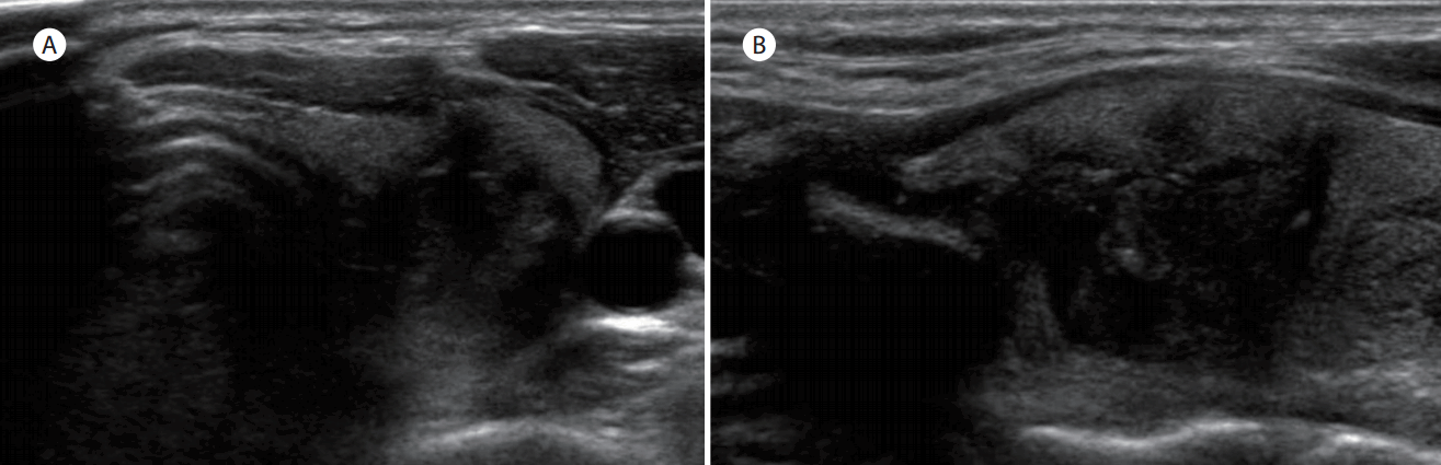
Case 14: solid variant papillary thyroid carcinoma in 52-year-old woman. Transverse (A) and longitudinal (B) sonograms shows a 0.8 cm taller than wide, spiculated, markedly hypoechoic nodule without calcification in right lobe of the thyroid.
Case 16: solid variant papillary thyroid carcinoma in 48-year-old woman. Transverse (A) and longitudinal (B) sonograms shows a 1.5 cm irregular, spiculated, marke hypoechoic mass with microcalcification in left lobe of the thyroid. Extrathyroidal extension of nodule was suspected on sonography.
고 찰
Nikiforov 등[8]은 고형 변이종 유두암의 유병률을 3% (20예/756예)로 보고하였으나, 최근 한국인을 대상으로 한 Chang 등[10]의 연구에서는 0.23% (14예/6,052예)를 보고하였다. 본 연구에서도 0.41% (17예/4,136예)로 Chang 등[10]과 비숫하였으며, Nikiforov 등[8]의 보고에 비해 유병률이 낮았다. 또한 고형 변이종 유두암은 방사선에 피폭된 병력이 있는 소아에서 잘 발생된다고 보고되어 있으나, Chang 등[10]의 연구에서는 방사선에 피폭된 병력을 가진 환자는 없었고 평균 연령은 48.2세였으며, 저자들의 경우도 방사선 피폭의 병력이 없었으며, 평균 연령이 49.2세였고, 13예(76.5%)는 45세 이상이어서 고형 변이종 유두암이 한국에서는 소아보다 성인에서 더 흔히 발생함을 알 수 있었다. 종양의 크기는 본 연구에서는 1.4 cm (범위 0.5-3.2 cm)로 상대적으로 종양의 크기가 작았으나, Giorgadze 등[11]의 연구에서는 평균 2.7 cm (범위 0.8-7.0 cm)였고, Nikiforov 등[8]은 평균 3.2 cm (범위 1.0-6.0 cm)로 종양이 저자들의 연구에 비해 컸다. Chang 등[10]의 경우도 평균 1.0 cm (범위 0.3-1.4 cm)로 대부분이 미세 유두암이었는데 이는 한국에서는 초음파검사에서 진단된 결절에 대한 초음파 유도하 세침흡인세포검사를 1 cm 이하의 작은 결절에도 시행함에 따른 결과로 해석할 수 있다. 그러나 저자들의 연구에서 결절의 크기가 작음에도 불구하고 12예(70.6%)에서 갑상선외 침범을 보였는데, 이는 Chang 등[10]의 50.0%와 Nikiforov 등[8]의 45.0%보다 훨씬 높았다. 또한 림프절 전이는 Nikiforov 등[8]은 55.0%를 보고하였고, Chang 등[10]의 35.7%를 보고하였는데, 본 연구에서는 10예(58.9%)로 Nikiforov 등[8]의 결과와 유사하여 고형 변이종 유두암은 주변조직의 침범이나 국소 림프절 전이가 잘됨을 알 수 있었다. Nikiforov 등[8]의 연구에서는 평균 18.7년의 경과관찰 중 수술 후 7년과 10년째에 원격전이로 인해 2예가 사망하였다고 보고하였는데, 저자들과 Chang 등[10]의 연구는 추적 기간이 5년 이하로 비교적 짧은 추적관찰 기간이어서 재발이나 원격전이 환자는 관찰되지 않았다.
갑상선 초음파검사는 다른 검사에 비해 방사선의 피폭이 없고, 검사가 단순하고 반복하기 쉬운 장점 외에도 갑상선 부피, 결절의 크기 측정, 갑상선 결절의 발견, 갑상선 결절의 특성 및 내부 성상, 전이성 림프절 진단 및 갑상선암의 주위 침윤 관찰, 갑상선암 수술 후 추적검사, Doppler 초음파를 이용하여 혈관 분포 확인, 초음파 유도하 세침흡인세포검사가 등의 장점이 있다. 초음파검사 소견 중 갑상선암과 연관된 것은 저에코성 음영과 미세석회화, 불규칙한 변연부, 결절의 앞뒤로 긴 모양, 피막을 침범한 경우, halo가 없음 등이 알려져 있다[11-15]. 이 중 저에코성 음영은 결절에서 흔히 관찰되고 악성보다는 양성 소견인 경우가 흔하여, 전경부의 피대근(strap muscle)의 에코와 유사한 음영을 보이는 현저한 저에코성 음영을 악성 종양의 지표로 이용하기도 한다[15]. Brito 등[16]은 초음파 관찰연구에 대한 메타 분석에서 갑상선 결절에 대한 악성을 예측하는데 갑상선 초음파의 민감도는 26-87%였고, 특이도는 40-93%를 보고하였으며, 여러 초음파 소견 중 앞뒤로 긴 모양이 악성을 나타내는 지표로 가장 승산비가 높다고 하였다.
유두암 변이종에 관한 초음파 특성에 관한 연구는 드물며, 키 큰 세포 변이종과 체모양-오디모양 변이종의 초음파 특징에 관한 연구가 보고되어 있다[17,18]. Choi 등[17]의 연구에서 8예의 키큰 세포 변이종은 초음파 소견상 현저한 저에코, 미세석회화와 갑상선 피막침범 등의 소견이 관찰된다고 하였으며, Chong 등[18]은 18예의 체모양-오디모양 변이종에 대한 초음파적 기준을 연구한 결과 17예는 불확실한 결절이었고 나머지 1예는 양성 결절로 진단되어 체모양-오디모양 변이종의 경우 초음파로 악성이 진단된 경우는 없었다고 한다. 고형 변이종 유두암의 초음파 특징에 관한 연구는 매우 드물며, Giorgadze 등[11]의 고형 변이종 유두암 13예에 대한 초음파 특징에 관한 연구가 보고되어 있다. Giorgadze 등[11]의 연구에서는 13예 중 5예(65.0%)가 경계가 분명한 결절이었고, 저에코 음영이 7예, 석회화는 2예에서 관찰되었다고 보고하였으나, 본 연구에서는 경계가 분명한 결절이 1예(5.9%)였고, 석회화도 10예(58.8%)에서 관찰되었다. 갑상선암을 예측하는 초음파 지표 중 앞뒤로 긴 모양은 Giorgadze 등[11]은 1예(7.7%)였으나, 저자들의 연구에서는 6예(35.3%)로 초음파 소견에서는 차이가 있었다. 이러한 차이는 Giorgadze 등[11]의 연구의 주된 목적이 초음파 특성을 보려 한 것이 아닌 것과 초음파 소견을 기술하는 방법의 차이와도 연관이 있을 것으로 추정되며, 향후 고형 변이종 유두암의 초음파 특징에 관한 연구가 더 흔히 보고가 되면 밝혀질 것으로 생각한다.
결론적으로 본 연구를 통하여 고형 변이종 유두암이 원격전이와 재발이 없어 공격적이지 않음을 알 수 있었다. 또한 고형 변이종 유두암은 초음파검사에서 앞뒤로 긴 모양, 현저한 저에코, 침상 변연부, 미세석회화 등의 악성을 시사하는 특징들을 보이는 경향이 있음을 알 수 있었다.