비장문의 림프절 종대를 동반한 원발성 비장 림프종
Primary Splenic Lymphoma with Splenic Hilar Lymphadenopathy
Article information
Abstract
71세 여자 환자가 경한 상복통과 비장종괴로 방문하였다. 복부 초음파 소견상 저에코의 단일 비장종괴 및 비장문 주위 림프절 종대가 관찰되었다. 전산화단층촬영 등 추가 복부 영상 검사 시행 후 정확한 진단 및 치료를 위해 비장절제술을 시행하였다. 조직 검사상 미만성거대B세포림프종으로 진단되었다. 비장종괴는 양성 병변이 많으나 비장문 주위 림프절 종대를 동반하는 단일 종괴성 병변인 경우 비장 림프종을 의심해 볼 수 있다. 또한 비장 림프종은 비장종괴 외 다양한 형태로 나타날 수도 있다.
Trans Abstract
Splenic lymphoma can occur as a primary or secondary malignancy, but the latter form is more common. Primary splenic lymphoma is defined as lymphomatous involvement of the spleen with or without splenic hilar lymphadenopathy. It can present as splenomegaly with homogenous echogenicity without a focal lesion, a diffuse infiltrative lesion with miliary lesions, multiple focal nodular lesions, or as a large solitary mass. A 71-year-old woman presented with mild upper abdominal pain and a splenic mass. We visualized a hypoechoic splenic mass with splenic hilar lymphadenopathy on abdominal ultrasound. The patient underwent surgery and was diagnosed with splenic diffuse large B-cell lymphoma.
서 론
비장의 원발성 림프종은 모든 림프종 환자의 1% 미만을 차지하는 매우 드문 질환이다[1]. 비장의 원발성 림프종은 일반적으로 말초 림프절 종대나 간 및 복강 내 림프종의 병변 없이 림프종이 비장과 비문 림프절에 국한된 경우를 말한다. 저자들은 상복부 통증과 비장종괴로 내원한 71세 여자 환자에서 복부 초음파 검사와 추가적인 영상 검사상 비장 림프종이 의심되어 시행한 수술적 치료 후 미만성거대B세포림프종(diffuse large B-cell lymphoma)으로 진단된 예를 보고한다.
증 례
71세 여자 환자가 개인병원 초음파상 비장종괴로 내원하였다. 특별한 과거력은 없었고 술은 거의 마시지 않았고 흡연력도 없었다. 환자는 경미한 상복부 통증을 호소하였고 압통은 없었다. 키는 153 cm, 몸무게는 60 kg이었다. 활력징후는 혈압 135/80 mmHg, 맥박 분당 70회, 호흡 분당 16회, 체온 37.0℃였다. 백혈구 8,300/mm3 (호중구 61%, 림프구 20%), 혈색소 14.3 g/dL, 혈소판 131,000/mm3, AST/ALT 13/20 U/L, ALP/γGTP 110/34 U/L, 총빌리루빈 0.8 mg/dL, amylase/lipase 60/37 U/L, BUN/Cr 20/1.1 mg/dL, HBs Ag/Ab(-/+), HCV Ab(-), CA 19-9 4.9 U/mL, CEA 1.0 ng/mL였다.
복부 초음파상 비교적 경계가 명확한 45 mm의 저에코의 단일 종괴가 관찰되었고 비문 주위의 저에코 림프절이 의심되었다(Fig. 1A and 1B). 도플러 초음파에서 특이한 혈관상은 보이지 않았고 비문 주위의 림프절이 의심되었다(Fig. 1C). 비장은 반우위로 촬영하였고 비장 크기는 정상이었고 단일 종괴였다.

(A) Abdominal ultrasonography of a 45-mm-sized hypoechoic mass in the spleen. (B, C) Gray-scale and color Doppler ultrasound reveal splenic hilar lymphadenopathy.
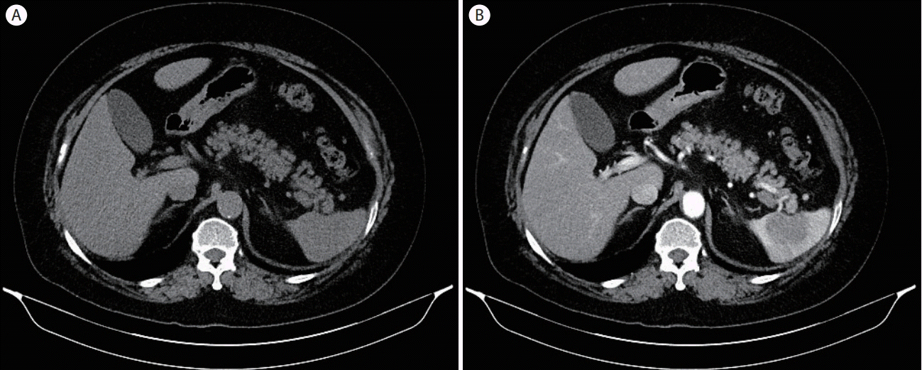
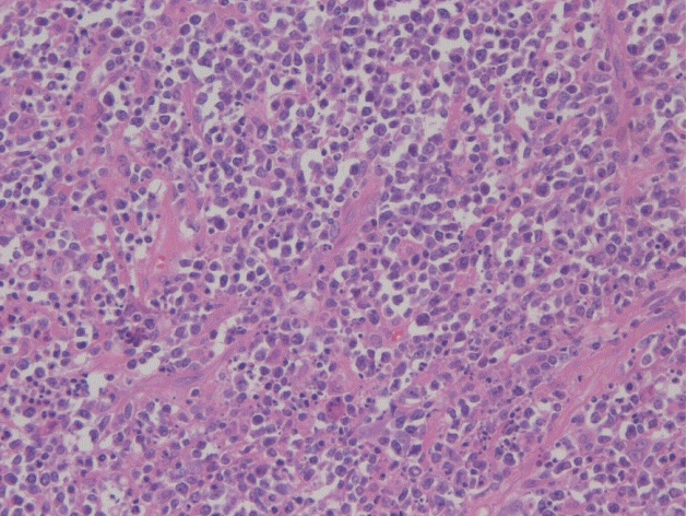
조영 전 컴퓨터단층촬영(computed tomography)에서 종괴는 비장과 유사한 소견이었고, 조영 후 비장종괴는 조영증강이 뚜렷하지 않았으며 비문주위 림프절이 관찰되었다(Fig. 2). T1 강조 자기공명영상(magnetic resonance imaging)에서 종괴는 비장과 유사한 소견이며, 조영 후 종괴의 주변이 조영증강되는 소견이었고 비문주위 림프절이 관찰되었다(Fig. 3). 여러 가지 복부 영상 검사 후 흔하지는 않지만 비문 주위 림프절 종대가 동반된 비장 림프종을 의심하여 수술을 시행하였다(Fig. 4). 수술 후 면역화학염색검사상 CD45, CD20 양성으로 나와서 미만성거대B 세포림프종으로 진단되었다(Figs. 5-7).

Abdominal computed tomography (CT). (A) Pre-contrast CT shows a suspicious low-density mass in the spleen. (B) Contrast-enhanced CT reveals splenic hilar lymphadenopathy without significant contrast enhancement of the splenic mass.

Magnetic resonance imaging (MRI). (A) T1-weighted MRI shows a suspicious low-signal mass in the spleen. (B) Enhanced MRI for the arterial phase demonstrates a peripheral-enhanced mass in the spleen and splenic hilar lymphadenopathy. (C) T2-weighted MRI reveals a mass signal similar to the spleen.
고 찰
비장의 림프종은 흔하지 않고 일차성 혹은 이차성이 있다[1]. 이차성 비장 림프종이 더 흔하며 원발성 비장 림프종은 드물다.
원발성 비장 림프종의 진단은 임상적 증상 외에 복부 초음파 검사나 복부 전산화단층촬영 등의 방사선 소견이 도움이 된다. 저음영의 비장종괴로 나타나는 경우가 있고 비장의 농양, 단순 낭포, 혈관종, 전이성 악성 종양, 과오종 등과의 감별 진단이 필요하다. 초음파 소견은 작은 경계를 가지는 결절, 5 mm 이하의 다수의 속립성 결절, 큰 저에코의 단발 종괴, 침윤형의 종괴가 없는 비장 종대 형태 등 다양하게 나타날 수 있다. 영상 검사에서 비장문 주위의 림프절 종대는 비장 림프종과 연관되는 소견으로 본 증례에서도 초음파, 컴퓨터단층촬영 등에서 관찰되었다.
원발성 비장 림프종의 가장 흔한 임상증상은 좌상복부 통증이며 체중 감소, 비장 비대, 피로감 등도 흔하며, 체중 감소, 열, 야간 발한 같은 B증상을 보이기도 한다[2]. 혈액학적으로 빈혈, 백혈구 감소, 혈소판 감소 및 적혈구 침강속도의 증가 소견을 보이는 경우도 있다.
비장을 침범하는 림프종의 병리학적 소견을 Ahmann 등[3]은 종괴가 없는 균질한 비장 비대, 속립성 종괴, 다발성 종괴, 큰 단발성 종괴의 네 가지로 분류하였는데, 특히 크기가 크면서 때때로 괴사를 동반한 종양의 경우 대세포형 림프종이 많다. 본 증례도 저에코의 큰 단발성 종괴로 미만성거대B세포림프종이었다.
원발성 비장 림프종의 치료로서 비장절제술이 확진 및 치료를 위해 효과적인 방법이며 국소방사선 치료나 복합화학요법 등을 병행하기도 한다[4].


