심초음파를 이용한 당뇨병성 심근병증의 진단
Abstract
Heart failure is common in the diabetic population, but while diabetic cardiomyopathy can cause heart failure, it is underestimated. Hyperglycemia, dyslipidemia and inflammation in diabetes are thought to play key roles in the pathogenesis of diabetic cardiomyopathy. Echocardiography has been the optimal noninvasive imaging test to evaluate the presence of cardiac dysfunction and it can provide clues to detect diabetic cardiomyopathy. This review discusses the echocardiographic findings of diabetic cardiomyopathy.
Keywords: Diabetic cardiomyopathy; Heart failure; Diabetes mellitus; Echocardiography
중심 단어: 당뇨병성 심근병증; 심부전; 당뇨병; 심초음파
서 론
당뇨병 환자에서 죽상동맥경화증, 허혈성 심질환, 고혈압 등이 동반되면서 심부전 발생이 발생할 수 있다. 또한 당뇨병성 심근병증(diabetic cardiomyopathy)도 당뇨병 환자에서 심부전의 원인이 될 수 있다[ 1]. 당뇨병성 심근병증은 1972년 심부전으로 사망한 당뇨병 환자 4명의 부검을 통해 처음 제기된 개념으로 다른 심부전의 원인은 없었다[ 2]. 이후 Framingham Heart Study를 통해 당뇨병이 있는 사람은 당뇨병이 없는 사람보다 남성은 2배, 여성은 5배 심부전이 더 많은 것이 확인되었다[ 3]. 당뇨병 환자에서 미세혈관장애, 대사이상, 세포 내 기관이상, 심장의 자율신경이상, 면역학적 부적응 반응 등이 관여하는 것으로 보인다[ 4]. 최근 당뇨병 치료제 중 sodium–glucose cotransporter 2 inhibitor (SGLT-2) 억제제는 심부전의 발생과 사망률을 감소시키는 것으로 나타나 심혈관질환, 특히 심부전을 동반한 당뇨병 환자에서 우선 투여를 고려하고 있다[ 5]. 따라서, 심초음파 등으로 당뇨병 환자에서 당뇨병성 심근병증에 의한 심부전의 초기 소견을 이해하면 치료에 도움이 될 것이다. 이에 저자들은 당뇨병성 심근병증에서 심초음파의 역할을 소개하고자 한다.
본 론
심초음파 소견
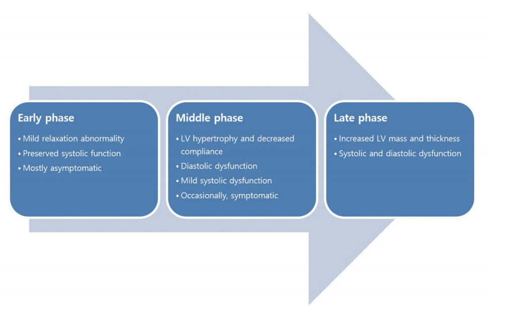
심초음파는 당뇨병성 심근병증을 진단함에 있어 표준 검사법이다. 또한 심장이상의 진행 정도나 치료의 반응을 평가하는 데도 유용한 도구이다[ 6]. 당뇨병성 심근병증은 구조적 이상과 기능적 이상을 동반하는데, 정도와 양상에 따라 병기를 구분하기도 한다[ 7] ( Fig. 1). 심초음파를 통해 발견할 수 있는 당뇨병성 심근병증의 대표적 이상 소견은 심근비후와 이완기능장애이다[ 8, 9]. 그러나, 확장성 심근병증과 유사한 형태(심내강의 확장과 수축기능장애)로 나타나는 경우도 있고[ 10], 초기에는 운동부하시에만 심초음파에서 이상 소견이 나타나기도 한다[ 11]. 즉, 이완기능장애와 심근비후가 당뇨병성 심근병증에서 특징적인 소견이지만 특이적이지는 않다[ 12].
심근비후와 심근질량의 증가
좌심실비후와 좌심실질량의 증가는 당뇨병에서 다양한 장기손상의 표현형 중 하나이다[ 13]. 심근이 두꺼워짐과 동시에 유순도가 감소(경직도가 증가)하여 이완기말에 좌심실로 유입되는 혈류량과 심박출량이 감소하고, 이로 인해 저관류와 울혈이 생겨 심부전의 증상과 징후가 나타날 수 있다[ 14]. 또한 제1형과 제2형 당뇨병 모두에서 좌심실비후를 동반한 심근병증이 발생할 수 있다[ 15]. 그러나 상대적으로 유병기간이 짧고 동반된 심혈관 질환의 유병률이 낮은 제1형 당뇨병 환자에서는 심근질량의 증가가 확연히 나타나지 않는다는 연구 결과도 있다[ 16, 17]. 좌심실질량의 증가는 당뇨병 환자에서 심부전을 일으키는 중요한 기여인자이면서 심혈관계 사망률의 예측인자이다. 따라서, 무증상 환자라도 선별검사를 통해 당뇨병성 심근병증을 조기에 진단하면 치료 계획을 수립하고 환자의 예후에 도움을 줄 수 있다[ 18]. 심근비후를 평가하는 첫 단계로 이면성 및 움직임 모드(motion mode, M-mode) 심초음파를 통해 심근벽의 두께를 측정한다[ 19]. 일반적으로 M-모드 심초음파를 통해 심실중격의 두께를 측정하며 이완기말에 11 mm를 넘으면 비후된 것으로 판단한다( Fig. 2). 이때 심근질량지수(left ventricular mass index)와 상대적 심실 두께(relative wall thickness)를 동시에 고려하면 좌심실의 재형성 양상(동심성 vs. 편심성, 단순 재형성 vs. 비대)을 구분할 수 있다[ 20]. 심근질량은 일반적으로 Devereux 공식(0.8 × 1.04 × [{(이완기말 좌심실 내경) + (이완기말 심실중격 두께) + (이완기말 좌심실 후벽 두께)} 3 – (이완기말 좌심실 내경) 3] + 0.6)를 이용하여 계산하고, 이를 체표면적으로 나누면 심근질량지수가 되며 남자에서는 < 115 g/m 2, 여자에서는 < 95 g/m 2을 정상으로 판단한다[ 21]. 또한 상대적 심실 두께는 ‘2 × (이완기말 좌심실 후벽 두께)/(이완기말 좌심실 내경)’의 공식을 이용하여 계산할 수 있고, > 0.42일 경우 동심성, 그렇지 않을 경우 편심성으로 본다[ 22]. 당뇨병성 심근병증의 좌심실비후는 동심성인 경우가 더 흔하며 상대적으로 더 특이적인 구조적 변화이다[ 18]. 이면성 심초음파에서 심근비후가 모호한 경우 3차원 영상을 이용하면 진단의 정확도를 높일 수 있다[ 23]. 심근비후와 별개로 심근의 간질 섬유화도 당뇨병성 심근병증 초기부터 발생할 수 있고 기능장애에 영향을 미친다[ 7, 24]. 심근의 섬유화는 일반적으로 이면성 심초음파에서 보이는 반향성으로 평가하지만, 최근 심근 조영자기공명영상을 통해 더 정확하고 정량적으로 평가하는 추세이다[ 25].
이완기능장애
심근비후는 심실의 유순도가 감소하는 수동적인 변화이다. 반대로 심근의 이완은 에너지를 소모하는 능동적인 과정이며, 결과적으로 두 요소 모두 이완기능장애 및 이완기말 심부전에 영향을 미친다[ 26]. 또한 심실비후 없이도 이완기능장애가 생길 수 있다[ 27]. 심장의 이완기능장애는 당뇨병성 심근병증의 초기부터 발생하는 기능적 변화로, 건강해 보이는 당뇨병 환자의 50%에서 발견되는 매우 흔한 이상이다[ 18, 28]. 이는 당뇨병과 당뇨병 전 단계 모두에서 발생할 수 있으며, 고령, 고혈압, 여성 당뇨병 환자에서 상대적으로 흔하다[ 29]. 또한 인슐린저항성이 높거나 혈당조절이 잘 되지 않을수록 발생률이 증가하며 중증도가 높다[ 30- 32]. 경도의 이완기능장애는 당뇨병 환자에서 예후에 미치는 영향이 크지 않지만, 장애가 진행되면 심부전의 발생률과 사망률이 증가한다[ 33]. 이완기능을 평가하기 위해 기본적으로 승모판유입혈류의 간헐파 도플러 영상에서 E파(초기 심실 충만을 반영)와 A파(심방 수축에 의한 후기 심실 충만을 반영)의 최고 속도, 초기 및 후기 이완기 최고 운동속도의 비(E/A ratio, E/A 비)를 측정한다( Fig. 3A). 색 도플러 M-모드로 승모판유입혈류에서 혈류전파속도(flow propagation velocity)를 측정해 이완기능장애를 평가할 수도 있지만, 전부하와 심박수의 영향을 받을 수 있으므로 널리 쓰이지는 않는다[ 34, 35]. 이완기능장애 초기에는 E/A 비가 역전되지만(< 0.8), 중기에는 좌심방 압력이 상승하면서 정상과 유사한 E/A 비(0.8-2.0)를 보이며 이를 가정상화(pseudonormalization)이라 한다[ 36]. 이때 발살바 술기(Valsalva maneuver)나 폐정맥혈류를 통해 정상과 구분할 수 있다. 후기에는 E파 속도가 더 증가하여 E/A 비도 증가한다(> 2.0). 이완기능의 정도를 평가하기 위해 추가적으로 좌심방용적지수(left atrial volume index)을 측정하여 활용한다( Fig. 3B and 3C). 또한 2016년 American Society of Echocardiography (ASE)/European Association of Cardiovascular Imaging (EACVI) 좌심실 이완기능 평가 가이드 라인에 삼첨판역류 최고 속도가 이완기능의 평가 지표로 추가되었으므로 함께 측정하도록 한다[ 36] ( Fig. 3D). 하지만 이 값만으로는 민감도와 특이도가 낮아 당뇨병성 심근병증을 진단하는 데 제한이 있다[ 37]. 예를 들어, E/A 비는 연령과 체액 용적 상태에 의존적이다[ 38]. 또한 검사자 간 차이가 생길 수 있고, 각 의존적(angle-dependent)이다. 이런 문제를 극복할 수 있는 가장 대표적인 기법이 조직 도플러 영상(tissue Doppler imaging) 기법이다. 이를 통해 전통적인 도플러 방법보다 특이적이면서 정량적으로 심근의 전반과 국소적인 분절의 수축 및 이완기능을 평가할 수 있다[ 39- 41] ( Fig. 3E and 3F). 그중에서도 E’ 속도는 이완기 때 승모판륜의 장축이동속도로 이완기능을 평가함에 있어 필수적이고, 이완기능장애 초기부터 감소되는 민감도가 높은 지표이다[ 30]. 제2형 당뇨병 환자에서 단순도플러 초음파만을 이용하여 진단할 때보다 조직 도플러 영상을 추가로 활용할 때 이완기능장애로 진단되는 비율이 50-74% 증가한다는 연구도 있다[ 30, 42]. E’ 속도는 가정상화 형태를 보이는 이완기능장애에서도 정상과 감별하는 데 유용하다. 또한 E/E’ 비를 통해 좌심실 이완기말 압력을 간접적이지만 정량적으로 알 수 있어 이완기능장애의 정도를 평가할 수 있다[ 36]. E/E’ (중격)이 15보다 크면 좌심실 이완기말 압력의 확연한 증가를 의미하며, 당뇨병 환자에서 심부전과 사망률이 증가한다[ 33]. 그러나, 이완기능을 평가할 때 심실특정 심근분획(중격 기저부나 외측벽 기저부)에서 E’ 속도를 측정하고 조직 도플러 영상이 조직의 수동적 움직임(예, 끌림[tethering])과 능동적 움직임을 구분하지 못하기 때문에 국소벽운동장애가 인접한 분획에 있을 경우 측정값이 영향을 받을 수 있다는 한계가 있다[ 43]. 2016년 ASE/ESCVI 가이드라인에 따르면 좌심실 구출률(heart failure with mid-range ejection fraction [HFmrEF])이 정상일 때 1. E/(중격과 자유벽의 E’ 평균) > 14, 2. 중격 E’ < 7 cm/s 또는 자유벽 E’ < 10 cm/s, 3. 삼첨판역류 최고 속도 > 2.8 m/s, 4. 좌심방용적지수 > 34 mL/m 2 중 3가지 또는 4가지를 만족할 때 이완기능 장애가 있으며, 2가지인 경우 모호하고, 0가지 또는 1가지인 경우 정상 이완기능으로 평가한다[ 36]. 그리고, 이완기능장애가 있다고 판단되는 경우 E/A 비, E파 최고 속도, E/(중격과 자유벽의 E’ 평균), 삼첨판역류 최고 속도, 좌심방용적지수 등을 이용하여 이완기능 장애의 정도를 구분한다.
수축기능장애
한 관찰 연구에 따르면 당뇨병성 심근병증 환자의 약 25%는 좌심실 구출률이 저하되었고, 약 75%는 유지되었다(이완기말심부전) [ 44]. 그러나, 당뇨병이 전반적인 좌심실 구출률 저하를 유발하는지에 대한 연구는 부족하다. 수축기능을 평가하는 대표적인 지표인 좌심실 구출률은 M-모드, 이면성, 3차원 영상 등 다양한 방법으로 측정할 수 있다. 측정한 좌심실 구출률을 바탕으로 심부전을 좌심실 구출률(HFmrEF, 40-50%), 좌심실 구출률(HFrEF, <40%)로 구분하며, 이에 따라 치료 방침이 달라진다[ 45]. 또한 조직 도플러 영상에서 수축기 최고 속도(S’)를 이용하여 수축기능을 평가할 수도 있다[ 46]. 당뇨병 환자는 동일한 좌심실 구출률이더라도 S’값이 감소되어 있다는 연구도 있다[ 47]. 최근 당뇨병 환자에서 심근의 변형(strain) 및 변형률(strain rate) 등을 심초음파나 자기공명영상을 이용하여 측정하는 연구가 활발하다. 변형은 심근의 지점 간 거리가 심주기에 따라 얼마나 변화하는지를 보여주는 값이며, 변형률은 그 변화 속도를 의미한다. 심장 관련 증상이 없고 좌심실 구출률이 보존되어 있는 초기 심근병증 환자에서도 변형이 감소되어 있을 수 있다. 따라서, 이를 측정하면 전반적인 좌심실기능의 변화를 매우 민감하게 진단할 수 있다. 그중에서 심초음파의 조직 도플러 영상으로 좌심실의 전반적인 종축변형(global longitudinal strain)을 측정하는 것이 가장 널리 쓰이는 방법이다. 무증상의 당뇨병 환자에서 좌심실의 전반적인 종축 변형은 당뇨병이 없는 사람에 비해 감소되어 있고, 이 경우 심혈관 사건 발생률이 증가한다[ 48, 49]. 반점 추적(speckle tracking) 기법은 전통적인 도플러나 조직 도플러 영상과 달리 각 분획을 직접 분석하므로 도플러 신호의 방향에 따라 생기는 오차가 없고, 전부하의 영향을 받지 않는다[ 43, 50]. 이로 인해 보다 직접적인 심근수축능의 평가가 가능하고, 검사자 간 오차도 발생하지 않는다. 또한 부수적으로 허혈성 심질환과 감별하고, 섬유화 유무를 파악하는 데 활용할 수 있다. 그러나, 변형은 심초음파 장비 사이에 표준화된 측정값이 없고, 정상과 이상을 구분하는 값의 경계에 대해 논란이 있어 아직 대중화되지 않았다. 요약하면 당뇨병성 심근병증에서 보이는 심초음파 소견은 다양하며 진단을 위한 단일 지표나 검사 기법이 없으므로 환자의 증상, 다양한 검사 결과를 종합적으로 판단하는 것이 필요하다. 심초음파를 통해 단순히 당뇨병성 심근병증을 진단하는 것뿐 아니라 혈당 조절, 동반질환의 영향으로 인한 변화를 추적 관찰하는 데에도 심초음파는 유용하다. 일례로 당뇨병 환자에서 당화혈색소가 2% 낮아지면 E’, 전반적인 종축변형값은 20% 이상 증가한다는 연구가 있다[ 51]. 마지막으로 허혈성 심질환 등 다른 원인으로 인하여 심초음파 이상 소견이 관찰되는 것은 아닌지 감별이 필요하다.
결 론
당뇨병 환자에서 주 사망 원인은 심혈관계 합병증으로 알려져 있다. 구체적으로 당뇨병에서 자율신경이상, 죽상동맥경화증에 의한 관상동맥질환, 심부전 발생 등이 동반된다. 당뇨병성 심근병증은 심부전의 발생 원인 중 하나로 고혈압, 허혈성 심질환, 심장판막질환 등 심부전의 다른 원인이 없을 때 고려해야 한다.
최근 당뇨병 치료제 중 SGLT-2 억제제 등은 심부전의 발생과 사망률을 줄이는 것으로 나타나고 있다. 당뇨병성 심근병증의심초음파 소견을 이해하면 적절한 치료 약물의 선택에 도움이 될 것이다.
Figure 1.
Progression of diabetic cardiomyopathy. LV, left ventricle. 
Figure 2.
Echocardiographic finding of diabetic cardiomyopathy– M mode image on the left parasternal window. The thickness of the interventricular septum and posterior wall of the left ventricle (LV) are increased. Calculated LV mass is 276.6 g and 138 g/m2 indexed by body surface area (reference value: < 115 g/m2 in male, and < 95 g/m2 in female) which indicates significant hypertrophy. Furthermore, relative wall thickness, which is calculated by the formula (2 x [posterior LV wall thickness]/[LV end-diastolic dimension]), is 0.62 (> 0.42 represents concentric hypertrophy). LVID, left ventricular internal dimension; LVPW, left ventricular posterior wall; IVS, interventricular septum; FS, fractional shortening; EF, ejection fraction. 
Figure 3.
Evaluation of diastolic function with various echocardiographic parameters. (A) Pulsed wave Doppler profile of mitral inflow. Peak velocity of E wave is increased, and E/A ratio is 2.3 which represents restrictive myocardium. (B, C) Measurement of volume of left atrium. Tracing atrial wall with Simpson’s method on apical 2- and 4-chamber view, volume of left atrium can be calculated. Dividing volume by body surface area, indexed volume of left atrium is derived (reference value: < 34 mL/m2). (D) Continuous wave Doppler pattern of tricuspid regurgitation. Peak velocity is 2.8 m/s, meeting 1 criterion of diastolic dysfunction. (E, F) Tissue Doppler image of interventricular septum (E) and lateral free wall (F). E’ velocity is decreased in both septum and free wall. E/E’ ratio is significantly increased by 32.4 (septum) and 23.8 (average of septum and free wall). This result represents increment of end-diastolic pressure of the left ventricle. Moreover, peak S’ velocity of septum (one of parameters demonstrating systolic function) is decreased. MV, mitral valve; DT, deceleration time; PHT, pressure half-time; DR, dynamic range; LA, left atrium; NTHI, native tissue harmonic imaging; 2D, two-dimensional. 
REFERENCES
2. Rubler S, Dlugash J, Yuceoglu YZ, Kumral T, Branwood AW, Grishman A. New type of cardiomyopathy associated with diabetic glomerulosclerosis. Am J Cardiol 1972;30:595–602.   3. Kannel WB, Hjortland M, Castelli WP. Role of diabetes in congestive heart failure: the Framingham study. Am J Cardiol 1974;34:29–34.   5. Iwakura K. Heart failure in patients with type 2 diabetes mellitus: assessment with echocardiography and effects of antihyperglycemic treatments. J Echocardiogr 2019;17:177–186.   6. Aneja A, Tang WH, Bansilal S, Garcia MJ, Farkouh ME. Diabetic cardiomyopathy: insights into pathogenesis, diagnostic challenges, and therapeutic options. Am J Med 2008;121:748–757.   7. Murtaza G, Virk HUH, Khalid M, et al. Diabetic cardiomyopathy-a comprehensive updated review. Prog Cardiovasc Dis 2019;62:315–326.   10. Paulus WJ, Tschöpe C, Sanderson JE, et al. How to diagnose diastolic heart failure: a consensus statement on the diagnosis of heart failure with normal left ventricular ejection fraction by the Heart Failure and Echocardiography Associations of the European Society of Cardiology. Eur Heart J 2007;28:2539–2550.   12. Fang ZY, Yuda S, Anderson V, Short L, Case C, Marwick TH. Echocardiographic detection of early diabetic myocardial disease. J Am Coll Cardiol 2003;41:611–617.   14. Park IB, Baik SH. Epidemiologic characteristics of diabetes mellitus in Korea: current status of diabetic patients using Korean Health Insurance Database. Korean Diabetes J 2009;33:357–362.  15. Teupe C, Rosak C. Diabetic cardiomyopathy and diastolic heart failure--difficulties with relaxation. Diabetes Res Clin Pract 2012;97:185–194.   16. Grandi AM, Piantanida E, Franzetti I, et al. Effect of glycemic control on left ventricular diastolic function in type 1 diabetes mellitus. Am J Cardiol 2006;97:71–76.   17. Palmieri V, Capaldo B, Russo C, et al. Left ventricular chamber and myocardial systolic function reserve in patients with type 1 diabetes mellitus: insight from traditional and Doppler tissue imaging echocardiography. J Am Soc Echocardiogr 2006;19:848–856.   19. Oh JK, Seward JB, Tajik AJ. The Echo Manual. 3rd Ed. London: Lippincott Williams & Wilkins,, 2006.
20. Ofstad AP, Urheim S, Dalen H, et al. Identification of a definite diabetic cardiomyopathy in type 2 diabetes by comprehensive echocardiographic evaluation: a cross-sectional comparison with non-diabetic weight-matched controls. J Diabetes 2015;7:779–790.   21. Devereux RB, Reicheck N. Echocardiographic determination of left ventricular mass in man. Anatomic validation of the method. Circulation 1977;55:613–618.   22. Lang RM, Badano LP, Mor-Avi V, et al. Recommendations for cardiac chamber quantification by echocardiography in adults: an update from the American Society of Echocardiography and the European Association of Cardiovascular Imaging. J Am Soc Echocardiogr 2015;28:1–39.e14.   23. Kusunose K, Kwon DH, Motoki H, Flamm SD, Marwick TH. Comparison of three-dimensional echocardiographic findings to those of magnetic resonance imaging for determination of left ventricular mass in patients with ischemic and non-ischemic cardiomyopathy. Am J Cardiol 2013;112:604–611.   24. Jellis C, Martin J, Narula J, Marwick TH. Assessment of nonischemic myocardial fibrosis. J Am Coll Cardiol 2010;56:89–97.   25. Di Bello V, Talarico L, Picano E, et al. Increased echodensity of myocardial wall in the diabetic heart: an ultrasound tissue characterization study. J Am Coll Cardiol 1995;25:1408–1415.   26. Kass DA, Bronzwaer JG, Paulus WJ. What mechanisms underlie diastolic dysfunction in heart failure? Circ Res 2004;94:1533–1542.   27. Schannwell CM, Schneppenheim M, Perings S, Plehn G, Strauer BE. Left ventricular diastolic dysfunction as an early manifestation of diabetic cardiomyopathy. Cardiology 2002;98:33–39.   28. Redfield MM, Jacobsen SJ, Burnett JC, Mahoney DW, Bailey KR, Rodeheffer RJ. Burden of systolic and diastolic ventricular dysfunction in the community: appreciating the scope of the heart failure epidemic. JAMA 2003;289:194–202.   29. Aguilar D, Deswal A, Ramasubbu K, Mann DL, Bozkurt B. Comparison of patients with heart failure and preserved left ventricular ejection fraction among those with versus without diabetes mellitus. Am J Cardiol 2010;105:373–377.   30. Boyer JK, Thanigaraj S, Schechtman KB, Pérez JE. Prevalence of ventricular diastolic dysfunction in asymptomatic, normotensive patients with diabetes mellitus. Am J Cardiol 2004;93:870–875.   32. Devereux RB, Roman MJ, Paranicas M, et al. Impact of diabetes on cardiac structure and function: the strong heart study. Circulation 2000;101:2271–2276.   33. From AM, Scott CJ, Chen HH. The development of heart failure in patients with diabetes mellitus and pre-clinical diastolic dysfunction a population-based study. J Am Coll Cardiol 2010;55:300–305.   34. Steine K, Stugaard M, Smiseth OA. Mechanisms of retarded apical filling in acute ischemic left ventricular failure. Circulation 1999;99:2048–2054.   35. Takatsuji H, Mikami T, Urasawa K, et al. A new approach for evaluation of left ventricular diastolic function: spatial and temporal analysis of left ventricular filling flow propagation by color M-mode Doppler echocardiography. J Am Coll Cardiol 1996;27:365–371.   36. Nagueh SF, Smiseth OA, Appleton CP, et al. Recommendations for the evaluation of left ventricular diastolic function by echocardiography: an update from the American Society of Echocardiography and the European Association of Cardiovascular Imaging. J Am Soc Echocardiogr 2016;29:277–314.   39. Miki T, Yuda S, Kouzu H, Miura T. Diabetic cardiomyopathy: pathophysiology and clinical features. Heart Fail Rev 2013;18:149–166.   40. Yu CM, Lin H, Ho PC, Yang H. Assessment of left and right ventricular systolic and diastolic synchronicity in normal subjects by tissue Doppler echocardiography and the effects of age and heart rate. Echocardiography 2003;20:19–27.   41. Yu CM, Sanderson JE, Marwick TH, Oh JK. Tissue Doppler imaging a new prognosticator for cardiovascular diseases. J Am Coll Cardiol 2007;49:1903–1914.   42. Shishehbor MH, Hoogwerf BH, Schoenhagen P, et al. Relation of hemoglobin A1c to left ventricular relaxation in patients with type 1 diabetes mellitus and without overt heart disease. Am J Cardiol 2003;91:1514–1517. A9.   46. Gulati VK, Katz WE, Follansbee WP, Gorcsan J 3rd. Mitral annular descent velocity by tissue Doppler echocardiography as an index of global left ventricular function. Am J Cardiol 1996;77:979–984.   47. Kosmala W, Kucharski W, Przewlocka-Kosmala M, Mazurek W. Comparison of left ventricular function by tissue Doppler imaging in patients with diabetes mellitus without systemic hypertension versus diabetes mellitus with systemic hypertension. Am J Cardiol 2004;94:395–399.   50. Greenberg NL, Firstenberg MS, Castro PL, et al. Doppler-derived myocardial systolic strain rate is a strong index of left ventricular contractility. Circulation 2002;105:99–105.   51. Leung M, Wong VW, Hudson M, Leung DY. Impact of improved glycemic control on cardiac function in type 2 diabetes mellitus. Circ Cardiovasc Imaging 2016;9:e003643.  
|
|


