만성 우상복부 불편감으로 나타난 담낭 편평상피세포암 1예
요약
74세 여성이 한 달간의 우상복부 불편감과 전신 쇠약감으로 시행한 복부 초음파검사에서 담낭벽의 불규칙한 비후와 담낭 내강으로 돌출된 종괴가 관찰되었다. 추가적인 영상검사에서 담낭암 혹은 황색 육아종 담낭염으로 추정되었고 수술 후 담낭 편평상피세포암으로 진단되었다. 담낭의 편평상피세포암은 담낭암의 드문 아형으로서 담낭 선암에 비하여 예후가 좋지 않다. 초음파 및 전산화단층촬영 등을 통한 조기 진단과 치료가 생존율 향상에 매우 중요하다.
중심 단어: 담낭암; 편평상피세포암; 초음파; 전산화단층촬영술
Abstract
A 74-year-old female patient was referred to our hospital with a one-month history of right upper abdominal discomfort and general weakness. Transabdominal ultrasonography conducted by a private clinic showed irregular wall thickening of the gallbladder and protruding mass into the gallbladder lumen. Computed tomography and magnetic resonance image in Wonkwang University Sanbon Hospital suggested gallbladder cancer or xanthogranulomatous cholecystitis. Extended cholecystectomy was performed. The finding of operation revealed gallbladder squamous cell carcinoma with mucosal destruction, emphysematous change of gallbladder and multiple gallbladder stones. Cancer invaded transverse colon, common bile duct, and hepatic artery. After the operation, the patient improved clinically, and is scheduled to treat adjuvant chemotherapy. The squamous cell carcinoma of the gallbladder is a rare histopathologic subtype of gallbladder cancer and has a highly proliferative character. Due to aggressive behavior, early detection and curative resection play an important role in survival benefit.
Keywords: Gallbladder cancer; Carcinoma, Squamous cell; Ultrasonography; Multidetector computed tomography
서 론
담낭암은 다른 암에 비하여 상대적으로 흔하지 않으며 조직학적으로 대부분(90%) 선암이다[ 1]. 담낭 편평상피세포암은 전체 담낭암 중 1-2%로 드물지만, 빠르게 성장하며 인접 기관을 침범하는 경향을 보여 예후가 좋지 않다[ 1- 3]. 그러나 담낭 편평상피세포암에 대한 병태생리는 연구가 많지 않아 잘 알려져 있지 않다. 이에 저자는 담석을 동반한 만성 담낭염 양상으로 나타난 담낭 편평상피세포암 증례를 경험하여 보고하는 바이다.
증 례
특이 과거력이 없는 74세 여성이 한 달간 지속된 우측 상복부 통증과 전신 쇠약을 주소로 개인의원에 방문하였다. 복부 초음파검사상 담낭벽의 불규칙한 비후와 담낭 내강으로 돌출된 종괴가 관찰되었다( Fig. 1). 본원으로 전원되어 시행한 이학적 검사상 우상복부 압통은 있었으나 murphy’s sign은 뚜렷하지 않았다. 혈액검사상 백혈구 11,500/uL (poly 76.3%), 혈색소 10.8/dL, 혈소판 379,600/uL, aspartate transaminase/alanine transaminase 25/22I U/L, 총 빌리루빈 0.49 mg/dL, blood urea nitrogen 23.6 mg/dL, creatinine 1.0 mg/dL, high sensitivity c-reactive protein 6.945 mg/dL, carbohydrate antigen 19-9 2.0 U/mL였다. 전산화단층촬영상 불규칙한 담낭벽의 비후와 담낭 주변 지방층에 선상음영을 동반한 기저부의 조영증강이 관찰되었고( Fig. 2), 자기공명영상촬영에서 담낭벽의 비후, 담낭 기저부의 점막 조영증강과 다발성 담석 그리고 담낭과 대망 주변부의 림프절이 확인되었다( Fig. 3). 영상검사를 바탕으로 담낭암(T3NxMx) 혹은 황색 육아종 담낭염(xanthogranulomatous cholecystitis)으로 추정 진단하고 개복수술을 결정하였다. 수술시 육안적으로 담낭은 매우 확장되어 있었으며, 표면은 딱딱하였고, 담낭의 기저부가 종괴형태로 2.5 cm 가량 두꺼워져 있었다. 두꺼워진 벽은 점막층 손상과 기종성 변화가 있었고 hartmann 주머니 내에 다발성 담석이 동반되었다. 확대담낭절제술을 시행하였고 병리학적 검사상 횡행대장의 장막, 총담관, 간동맥을 침범한 중등도 분화도(G2)의 담낭 편평상피세포암으로 진단되었다( Fig. 4). 하지만 담낭 주변부의 림프절은 암의 침범을 보이지 않는 것으로 확인하여 TNM 병기상 T4N0M0로 최종 진단되었다. 수술 후 환자는 임상적으로 호전되어 퇴원하였고 향후 보조 항암치료를 할 예정이다.
고 찰
담낭 편평상피세포암은 임상양상, 이학적 검사상 소견, 담석 동반 유무 등의 측면에서 볼 때 다른 아형의 담낭암과 큰 차이는 없다[ 2, 3]. 우상복부 혹은 상복부 불편감이 흔한 증상이며 황달, 발열 등도 나타날 수 있다[ 3]. 다른 아형의 담낭암과 같이 담낭 편평상피세포암은 여성에게서 호발하며(남:여 = 3.8:1), 발생 평균 나이는 60대이다[ 2- 4]. 담낭 편평상피세포암의 호발 부위는 기저부(39%)이며[ 5], 질환의 희귀성으로 인해 위험인자가 정립되어 있지는 않으나 담석을 위험인자로 보는 견해도 있다[ 2]. 본 증례의 환자도 기저부에서 발생하였고 다발성 담석이 동반되었다. 담낭 선암에 비하여 높은 증식성을 보이는 특징으로 인해 많은 환자들에서 담낭 선암보다 더 진행된 병기로 진단되는 경우가 많다[ 2, 3]. 담낭 편평상피세포암은 인접 기관의 침범이 빈번하며 가장 흔하게 침범하는 기관은 간이고 이외에 담관이나 소화관, 혈관들의 침범도 보고되고 있다[ 3, 6]. 초기 검사에서 간기능검사, 암표지자검사 및 영상검사 등을 시행해야 하고, 황달은 담낭암의 불량한 예후인자로 알려져 있다. 고정된 용종형 종괴, 국소 담낭벽 비후가 동반된 담낭 내강내 종괴 돌출, 담낭의 폭 증가 그리고 담낭과 간 표면 사이 경계면 소실 등이 복부 초음파에서 관찰된다면 담낭암의 가능성을 시사한다[ 7, 8]. 전산화단층촬영, 자기공명영상촬영은 림프절 전이, 인접기관 침범, 원격 전이를 평가하는 데 도움이 되고 황달이 있는 경우 자기공명담췌관조영술도 고려해야 한다[ 8]. 담낭 편평상피세포암은 기존에 담낭내 편평상피화생이 된 장소에서 발생하는 것으로 알려져 있다. 이상각화를 포함하는 광범위한 각화와 종양내 호산구 침윤 등이 담낭 편평상피세포암의 조직학적 특징이다[ 2]. 담낭 편평상피세포암은 담낭 선암에 비하여 예후가 좋지 않다. Roa 등[ 2]의 연구에 의하면 중위수 생존 기간(median survival time)을 4개월로 보고하고 있다. Henson 등[ 4]의 연구에서는 2년 생존율을 9%로 보고하였고, Kim 등[ 3]의 연구에서는 1년 생존율이 담낭 선암에 비하여 현저히 낮음을 보고하였다(18.8% vs. 87.3%). 담낭 편평상피세포암은 근치적 치료를 위해서는 절제 변연 음성을 동반한 완전한 절제가 필요하다[ 3, 6]. 최근 수술 후 보조화학요법과 방사선치료에 대한 몇몇 시도가 있었다. Bourmèche 등[ 9]의 연구에 의하면 5 fluorouracil과 cisplatin을 방사선치료와 병합하여 완전 관해를 보고한 바 있다. 담낭 편평상피세포암은 담낭암의 드문 아형 중 하나로서 선암에 비하여 훨씬 진행된 병기로 진단되는 경우가 많고 예후가 좋지 않다. 그러므로 조기 진단과 근치적 절제만이 생존율 개선에 매우 중요하다고 할 수 있다. 향후 담낭 편평상피세포암에 대한 병태생리 및 치료법 등에 대한 전향적인 연구가 필요할 것으로 생각된다.
Figure 1.
Transabdominal ultrasound showed irregular wall thickening of the gallbladder and protruding mass into the gallbladder lumen (arrow). 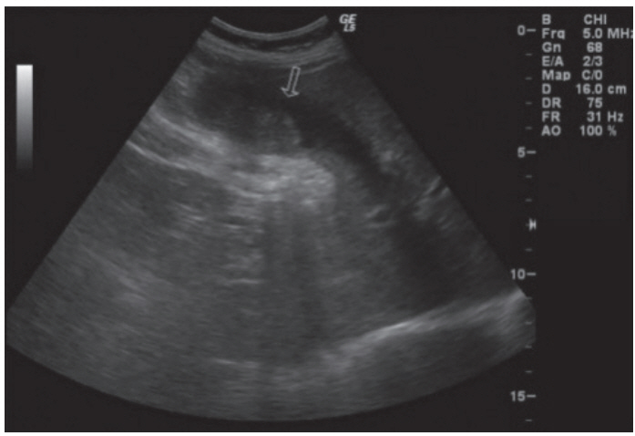
Figure 2.
Computed tomography revealed irregular wall thickening and increased wall enhancement at gallbladder fundus with pericholecystic fat stranding. 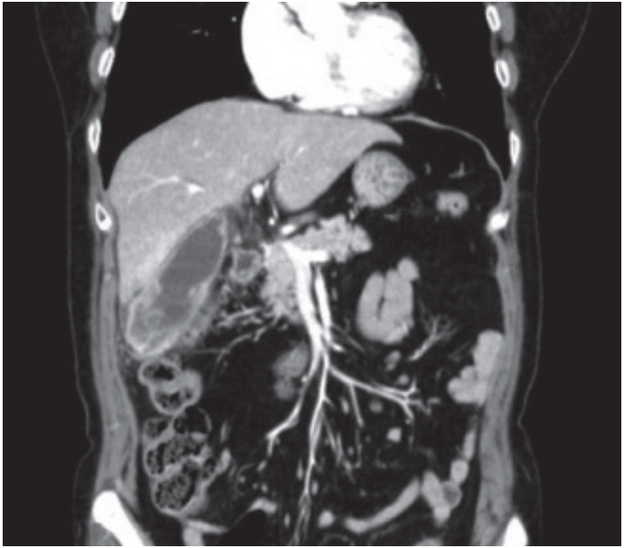
Figure 3.
Magnetic resonance imaging of the gallbladder revealed increased mucosal enhancement at gallbladder fundus and multiple stones in the gallbladder fundus and cystic duct. 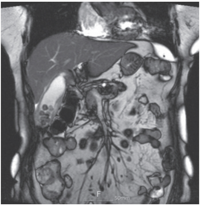
Figure 4.
The macroscopic finding showed an irregular ill-defined mass with hard consistency and necrotic surface in the gallbladder fundus. 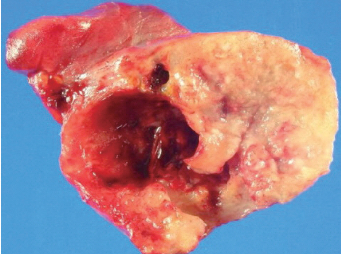
REFERENCES
1. Levy AD, Murakata LA, Rohrmann CA Jr. Gallbladder carcinoma: radiologic-pathologic correlation. Radiographics 2001;21:295–314. questionnaire, 549-555.   3. Kim WS, Jang KT, Choi DW, et al. Clinicopathologic analysis of adenosquamous/squamous cell carcinoma of the gallbladder. J Surg Oncol 2011;103:239–242. 4. Henson DE, Albores-Saavedra J, Corle D. Carcinoma of the gallbladder. Histologic types, stage of disease, grade, and survival rates. Cancer 1992;70:1493–1497. 5. Andrea C, Francesco C. Squamous-cell and non-squamouscell carcinomas of the gallbladder have different risk factors. Lancet Oncol 2003;4:393–394. 6. Chan KM, Yu MC, Lee WC, Jan YY, Chen MF. Adenosquamous/squamous cell carcinoma of the gallbladder. J Surg Oncol 2007;95:129–134. 7. Misra S, Chaturvedi A, Misra NC, Sharma ID. Carcinoma of the gallbladder. Lancet Oncol 2003;4:167–176. 8. Wang JH, Liu BJ, Xu HX, et al. Clinical, pathological and sonographic characteristics of unexpected gallbladder carcinoma. Int J Clin Exp Med 2015;8:11109–11116.  9. Bourmèche M, Ben Salah H, Kallel M, et al. A long survival after the treatment of a squamous cell carcinoma of the gallbladder. Cancer Radiother 2013;17:58–61.
|
|


